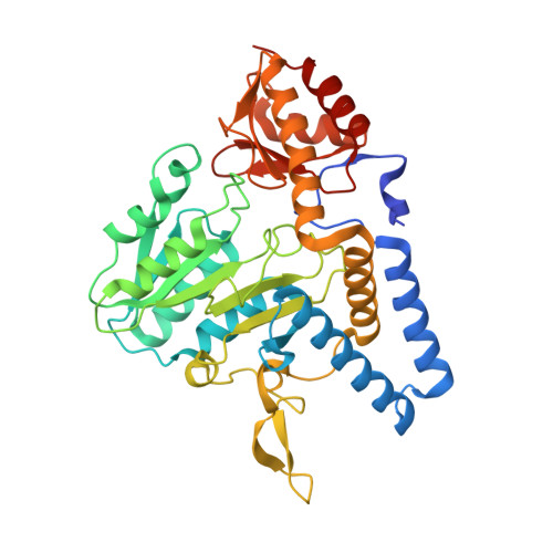
1ELQ

CRYSTAL STRUCTURE OF THE CYSTINE C-S LYASE C-DES


Experimental Data Snapshot

  • Method: X-RAY DIFFRACTION
  • Resolution: 1.80 Å
  • R-Value Free: 0.249 
  • R-Value Work: 0.198 
  • R-Value Observed: 0.201 

wwPDB Validation   3D Report Full Report


Ligand Structure Quality Assessment 


This is version 1.3 of the entry. See complete history


Literature

Crystal structure of the cystine C-S lyase from Synechocystis: stabilization of cysteine persulfide for FeS cluster biosynthesis.

Clausen, T.Kaiser, J.T.Steegborn, C.Huber, R.Kessler, D.

(2000) Proc Natl Acad Sci U S A 97: 3856-3861

  • DOI: https://doi.org/10.1073/pnas.97.8.3856
  • Primary Citation of Related Structures:  
    1ELQ, 1ELU

  • PubMed Abstract: 

    FeS clusters are versatile cofactors of a variety of proteins, but the mechanisms of their biosynthesis are still unknown. The cystine C-S lyase from Synechocystis has been identified as a participant in ferredoxin FeS cluster formation. Herein, we report on the crystal structure of the lyase and of a complex with the reaction products of cystine cleavage at 1.8- and 1.55-A resolution, respectively. The sulfur-containing product was unequivocally identified as cysteine persulfide. The reactive persulfide group is fixed by a hydrogen bond to His-114 in the center of a hydrophobic pocket and is thereby shielded from the solvent. Binding and stabilization of the cysteine persulfide represent an alternative to the generation of a protein-bound persulfide by NifS-like proteins and point to the general importance of persulfidic compounds for FeS cluster assembly.


  • Organizational Affiliation

    Max-Planck-Institut für Biochemie, Abteilung Strukturforschung, Am Klopferspitz 18a, D-82152 Martinsried, Germany. clausen@biochem.mpg.de


Macromolecules
Find similar proteins by:  (by identity cutoff)  |  3D Structure
Entity ID: 1
MoleculeChains Sequence LengthOrganismDetailsImage
L-CYSTEINE/L-CYSTINE C-S LYASE
A, B
390Synechocystis sp. PCC 6714Mutation(s): 8 
UniProt
Find proteins for Q9ZHG9 (Synechocystis sp. (strain PCC 6714))
Explore Q9ZHG9 
Go to UniProtKB:  Q9ZHG9
Entity Groups  
Sequence Clusters30% Identity50% Identity70% Identity90% Identity95% Identity100% Identity
UniProt GroupQ9ZHG9
Sequence Annotations
Expand
  • Reference Sequence
Experimental Data & Validation

Experimental Data

  • Method: X-RAY DIFFRACTION
  • Resolution: 1.80 Å
  • R-Value Free: 0.249 
  • R-Value Work: 0.198 
  • R-Value Observed: 0.201 
  • Space Group: P 21 21 21
Unit Cell:
Length ( Å )Angle ( ˚ )
a = 62.4α = 90
b = 65.4β = 90
c = 170.1γ = 90
Software Package:
Software NamePurpose
SHARPphasing
CNSrefinement
DENZOdata reduction
CCP4data scaling

Structure Validation

View Full Validation Report


Ligand Structure Quality Assessment 


Entry History 

Deposition Data

Revision History  (Full details and data files)

  • Version 1.0: 2000-04-19
    Type: Initial release
  • Version 1.1: 2008-04-27
    Changes: Version format compliance
  • Version 1.2: 2011-07-13
    Changes: Source and taxonomy, Version format compliance
  • Version 1.3: 2021-11-03
    Changes: Data collection, Database references, Derived calculations