Structures of thermolabile mutants of human glutathione transferase P1-1.
Rossjohn, J., McKinstry, W.J., Oakley, A.J., Parker, M.W., Stenberg, G., Mannervik, B., Dragani, B., Cocco, R., Aceto, A.(2000) J Mol Biol 302: 295-302
- PubMed: 10970734 Search on PubMed
- DOI: https://doi.org/10.1006/jmbi.2000.4054
- Primary Citation Related Structures:
1EOG, 1EOH - PubMed Abstract:
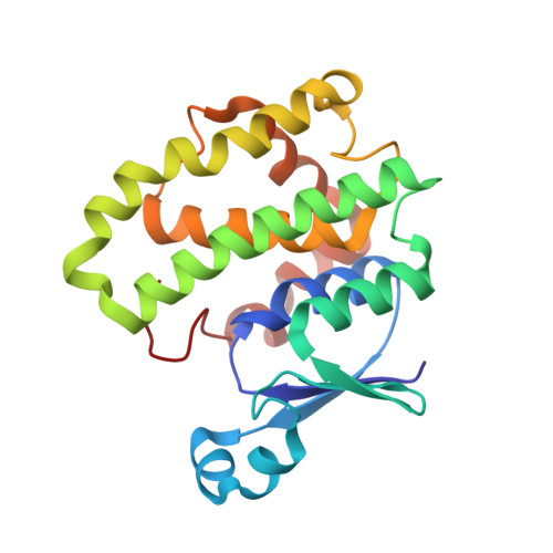
An N-capping box motif (Ser/Thr-Xaa-Xaa-Asp) is strictly conserved at the beginning of helix alpha6 in the core of virtually all glutathione transferases (GST) and GST-related proteins. It has been demonstrated that this local motif is important in determining the alpha-helical propensity of the isolated alpha6-peptide and plays a crucial role in the folding and stability of GSTs. Its removal by site-directed mutagenesis generated temperature-sensitive folding mutants unable to refold at physiological temperature (37 degrees C). In the present work, variants of human GSTP1-1 (S150A and D153A), in which the capping residues have been substituted by alanine, have been generated and purified for structural analysis. Thus, for the first time, temperature-sensitive folding mutants of an enzyme, expressed at a permissive temperature, have been crystallized and their three-dimensional structures determined by X-ray crystallography. The crystal structures of human pi class GST temperature-sensitive mutants provide a basis for understanding the structural origin of the dramatic effects observed on the overall stability of the enzyme at higher temperatures upon single substitution of a capping residue.
- The Ian Potter Foundation Protein Crystallography Laboratory, St. Vincent's Institute of Medical Research, Fitzroy, Victoria, 3065, Australia.
Organizational Affiliation: