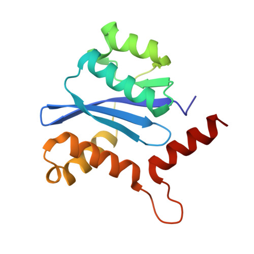
1EXQ

CRYSTAL STRUCTURE OF THE HIV-1 INTEGRASE CATALYTIC CORE DOMAIN


Experimental Data Snapshot

  • Method: X-RAY DIFFRACTION
  • Resolution: 1.60 Å
  • R-Value Free: 0.271 
  • R-Value Work: 0.253 
  • R-Value Observed: 0.253 

wwPDB Validation   3D Report Full Report


This is version 1.8 of the entry. See complete history


Literature

Crystal structure of the HIV-1 integrase catalytic core and C-terminal domains: a model for viral DNA binding.

Chen, J.C.Krucinski, J.Miercke, L.J.Finer-Moore, J.S.Tang, A.H.Leavitt, A.D.Stroud, R.M.

(2000) Proc Natl Acad Sci U S A 97: 8233-8238

  • DOI: https://doi.org/10.1073/pnas.150220297
  • Primary Citation of Related Structures:  
    1EX4, 1EXQ

  • PubMed Abstract: 

    Insolubility of full-length HIV-1 integrase (IN) limited previous structure analyses to individual domains. By introducing five point mutations, we engineered a more soluble IN that allowed us to generate multidomain HIV-1 IN crystals. The first multidomain HIV-1 IN structure is reported. It incorporates the catalytic core and C-terminal domains (residues 52-288). The structure resolved to 2.8 A is a Y-shaped dimer. Within the dimer, the catalytic core domains form the only dimer interface, and the C-terminal domains are located 55 A apart. A 26-aa alpha-helix, alpha6, links the C-terminal domain to the catalytic core. A kink in one of the two alpha6 helices occurs near a known proteolytic site, suggesting that it may act as a flexible elbow to reorient the domains during the integration process. Two proteins that bind DNA in a sequence-independent manner are structurally homologous to the HIV-1 IN C-terminal domain, suggesting a similar protein-DNA interaction in which the IN C-terminal domain may serve to bind, bend, and orient viral DNA during integration. A strip of positively charged amino acids contributed by both monomers emerges from each active site of the dimer, suggesting a minimally dimeric platform for binding each viral DNA end. The crystal structure of the isolated catalytic core domain (residues 52-210), independently determined at 1.6-A resolution, is identical to the core domain within the two-domain 52-288 structure.


  • Organizational Affiliation

    Departments of Biochemistry and Biophysics, Laboratory Medicine, and Internal Medicine, University of California, San Francisco, CA 94143, USA. stroud@msg.ucsf.edu


Macromolecules
Find similar proteins by:  (by identity cutoff)  |  3D Structure
Entity ID: 1
MoleculeChains Sequence LengthOrganismDetailsImage
POL POLYPROTEIN
A, B
154Human immunodeficiency virus 1Mutation(s): 4 
UniProt
Find proteins for P04585 (Human immunodeficiency virus type 1 group M subtype B (isolate HXB2))
Explore P04585 
Go to UniProtKB:  P04585
Entity Groups  
Sequence Clusters30% Identity50% Identity70% Identity90% Identity95% Identity100% Identity
UniProt GroupP04585
Sequence Annotations
Expand
  • Reference Sequence
Small Molecules
Ligands 3 Unique
IDChains Name / Formula / InChI Key2D Diagram3D Interactions
CD
Query on CD

Download Ideal Coordinates CCD File 
C [auth A],
D [auth A],
G [auth B],
H [auth B]
CADMIUM ION
Cd
WLZRMCYVCSSEQC-UHFFFAOYSA-N
SO4
Query on SO4

Download Ideal Coordinates CCD File 
F [auth A],
J [auth B],
K [auth B],
L [auth B]
SULFATE ION
O4 S
QAOWNCQODCNURD-UHFFFAOYSA-L
CL
Query on CL

Download Ideal Coordinates CCD File 
E [auth A],
I [auth B]
CHLORIDE ION
Cl
VEXZGXHMUGYJMC-UHFFFAOYSA-M
Experimental Data & Validation

Experimental Data

  • Method: X-RAY DIFFRACTION
  • Resolution: 1.60 Å
  • R-Value Free: 0.271 
  • R-Value Work: 0.253 
  • R-Value Observed: 0.253 
  • Space Group: P 32
Unit Cell:
Length ( Å )Angle ( ˚ )
a = 48.886α = 90
b = 48.886β = 90
c = 103.644γ = 120
Software Package:
Software NamePurpose
CNSrefinement
MAR345data collection
SCALEPACKdata scaling
CNSphasing

Structure Validation

View Full Validation Report


Entry History 

Deposition Data

Revision History  (Full details and data files)

  • Version 1.0: 2000-11-03
    Type: Initial release
  • Version 1.1: 2008-04-27
    Changes: Version format compliance
  • Version 1.2: 2011-07-13
    Changes: Version format compliance
  • Version 1.3: 2017-02-01
    Changes: Structure summary
  • Version 1.4: 2017-10-04
    Changes: Refinement description
  • Version 1.5: 2018-01-24
    Changes: Structure summary
  • Version 1.6: 2018-01-31
    Changes: Experimental preparation
  • Version 1.7: 2021-11-03
    Changes: Database references, Derived calculations
  • Version 1.8: 2024-02-07
    Changes: Data collection