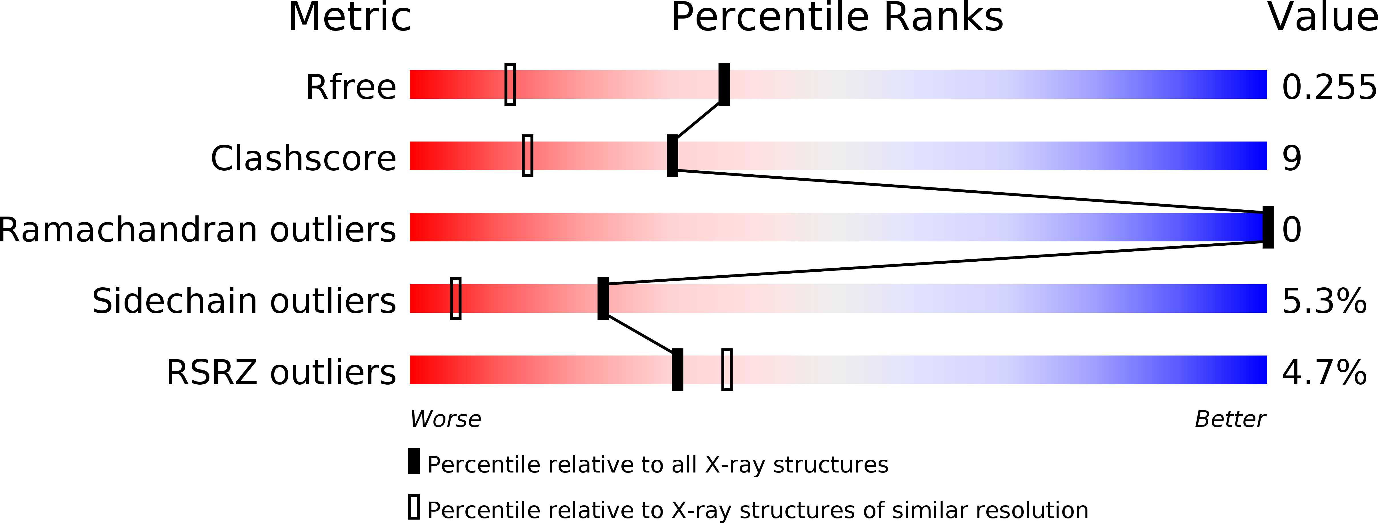
1F32

CRYSTAL STRUCTURE OF ASCARIS PEPSIN INHIBITOR-3


Experimental Data Snapshot

  • Method: X-RAY DIFFRACTION
  • Resolution: 1.75 Å
  • R-Value Free: 
    0.255 (Depositor), 0.250 (DCC) 
  • R-Value Work: 
    0.211 (Depositor), 0.210 (DCC) 

wwPDB Validation   3D Report Full Report


This is version 1.4 of the entry. See complete history


Literature

Structural basis for the inhibition of porcine pepsin by Ascaris pepsin inhibitor-3.

Ng, K.K.Petersen, J.F.Cherney, M.M.Garen, C.Zalatoris, J.J.Rao-Naik, C.Dunn, B.M.Martzen, M.R.Peanasky, R.J.James, M.N.

(2000) Nat Struct Biol 7: 653-657

  • DOI: https://doi.org/10.1038/77950
  • Primary Citation of Related Structures:  
    1F32, 1F34

  • PubMed Abstract: 

    The three-dimensional structures of pepsin inhibitor-3 (PI-3) from Ascaris suum and of the complex between PI-3 and porcine pepsin at 1. 75 A and 2.45 A resolution, respectively, have revealed the mechanism of aspartic protease inhibition by this unique inhibitor. PI-3 has a new fold consisting of two domains, each comprising an antiparallel beta-sheet flanked by an alpha-helix. In the enzyme-inhibitor complex, the N-terminal beta-strand of PI-3 pairs with one strand of the 'active site flap' (residues 70-82) of pepsin, thus forming an eight-stranded beta-sheet that spans the two proteins. PI-3 has a novel mode of inhibition, using its N-terminal residues to occupy and therefore block the first three binding pockets in pepsin for substrate residues C-terminal to the scissile bond (S1'-S3'). The molecular structure of the pepsin-PI-3 complex suggests new avenues for the rational design of proteinaceous aspartic proteinase inhibitors.


  • Organizational Affiliation

    MRC of Canada, Group in Protein Structure and Function, Department of Biochemistry, University of Alberta, Edmonton, Alberta T6G 2H7, Canada.


Macromolecules
Find similar proteins by:  (by identity cutoff)  |  3D Structure
Entity ID: 1
MoleculeChains Sequence LengthOrganismDetailsImage
MAJOR PEPSIN INHIBITOR PI-3149Ascaris suumMutation(s): 0 
UniProt
Find proteins for P19400 (Ascaris suum)
Explore P19400 
Go to UniProtKB:  P19400
Entity Groups  
Sequence Clusters30% Identity50% Identity70% Identity90% Identity95% Identity100% Identity
UniProt GroupP19400
Sequence Annotations
Expand
  • Reference Sequence
Experimental Data & Validation

Experimental Data

  • Method: X-RAY DIFFRACTION
  • Resolution: 1.75 Å
  • R-Value Free:  0.255 (Depositor), 0.250 (DCC) 
  • R-Value Work:  0.211 (Depositor), 0.210 (DCC) 
Space Group: P 1 21 1
Unit Cell:
Length ( Å )Angle ( ˚ )
a = 30.455α = 90
b = 72.717β = 101.03
c = 31.137γ = 90
Software Package:
Software NamePurpose
DENZOdata reduction
SCALEPACKdata scaling
SOLVEphasing
CNSrefinement

Structure Validation

View Full Validation Report


Entry History 

Deposition Data

Revision History  (Full details and data files)

  • Version 1.0: 2001-02-01
    Type: Initial release
  • Version 1.1: 2008-04-27
    Changes: Version format compliance
  • Version 1.2: 2011-07-13
    Changes: Version format compliance
  • Version 1.3: 2018-04-04
    Changes: Data collection
  • Version 1.4: 2024-11-13
    Changes: Data collection, Database references, Structure summary