1FQ3 | pdb_00001fq3

CRYSTAL STRUCTURE OF HUMAN GRANZYME B


Experimental Data Snapshot

  • Method: X-RAY DIFFRACTION
  • Resolution: 3.10 Å

Starting Model: experimental
View more details

wwPDB Validation 3D Report Full Report

Validation slider image for 1FQ3

This is version 2.2 of the entry. See complete history

Literature

Crystal structure of the caspase activator human granzyme B, a proteinase highly specific for an Asp-P1 residue.

Estebanez-Perpina, E.Fuentes-Prior, P.Belorgey, D.Braun, M.Kiefersauer, R.Maskos, K.Huber, R.Rubin, H.Bode, W.

(2000) Biol Chem 381: 1203-1214

  • DOI: https://doi.org/10.1515/BC.2000.148
  • Primary Citation Related Structures: 
    1FQ3

  • PubMed Abstract: 

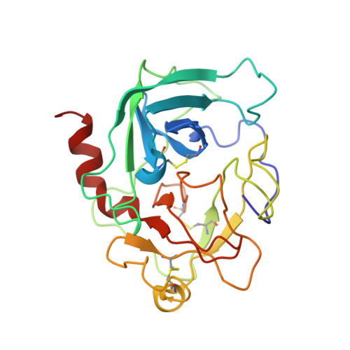
    Granzyme B is the prototypic member of the granzymes, a family of trypsin-like serine proteinases localized in the dense cytoplasmic granules of activated natural killer cells and cytotoxic T lymphocytes. Granzyme B directly triggers apoptosis in target cells by activating the caspase pathway, and has been implicated in the etiology of rheumatoid arthritis. Human granzyme B expressed in a baculovirus system has been crystallized without inhibitor and its structure has been determined to 3.1 A resolution, after considerably improving the diffraction power of the crystals by controlled humidity changes. The granzyme B structure reveals an overall fold similar to that found in cathepsin G and human chymase. The guanidinium group of Arg226, anchored at the back of the S1-specificity pocket, can form a salt bridge with the P1-Asp side chain of a bound peptide substrate. The architecture of the substrate binding site of granzyme B appears to be designed to accommodate and cleave hexapeptides such as the sequence Ile-Glu-Thr-Asp-/Ser-Gly present in the activation site of pro-caspase-3, a proven physiological substrate of granzyme B. These granzyme B crystals, with fully accessible active sites, are well suited for soaking with small synthetic inhibitors that might be used for a treatment of chronic inflammatory disorders.


  • Organizational Affiliation
    • Abteilung für Strukturforschung, Max-Planck-Institut für Biochemie, Planegg-Martinsried, Germany.

Macromolecule Content 

  • Total Structure Weight: 52.47 kDa 
  • Atom Count: 3,670 
  • Modeled Residue Count: 454 
  • Deposited Residue Count: 454 
  • Unique protein chains: 1

Macromolecules

Find similar proteins by:|  3D Structure
Entity ID: 1
MoleculeChains  Sequence LengthOrganismDetailsImage
GRANZYME B
A, B
227Homo sapiensMutation(s): 0 
EC: 3.4.21.79
UniProt & NIH Common Fund Data Resources
Find proteins for P10144 (Homo sapiens)
Explore P10144 
Go to UniProtKB:  P10144
PHAROS:  P10144
GTEx:  ENSG00000100453 
Entity Groups
Sequence Clusters30% Identity50% Identity70% Identity90% Identity95% Identity100% Identity
UniProt GroupP10144
Glycosylation
Glycosylation Sites: 1Go to GlyGen: P10144-1
Sequence Annotations
Expand
Reference Sequence

Oligosaccharides

Help  
Entity ID: 2
MoleculeChains Length2D Diagram GlycosylationD Interactions
beta-D-mannopyranose-(1-4)-2-acetamido-2-deoxy-beta-D-glucopyranose-(1-4)-2-acetamido-2-deoxy-beta-D-glucopyranose
C, D
3N-Glycosylation
Glycosylation Resources
GlyTouCan: G15407YE
GlyCosmos: G15407YE
GlyGen: G15407YE

Small Molecules

Experimental Data & Validation

Experimental Data

  • Method: X-RAY DIFFRACTION
  • Resolution: 3.10 Å
Space Group: P 21 21 21
Unit Cell:
Length ( Å )Angle ( ˚ )
a = 41.74α = 90
b = 114.31β = 90
c = 135.52γ = 90
Software Package:
Software NamePurpose
MOSFLMdata reduction
SCALAdata scaling
AMoREphasing
X-PLORrefinement
CCP4data scaling

Structure Validation

View Full Validation Report


Entry History 

Deposition Data

Revision History  (Full details and data files)

  • Version 1.0: 2001-01-31
    Type: Initial release
  • Version 1.1: 2008-04-27
    Changes: Version format compliance
  • Version 1.2: 2011-07-13
    Changes: Non-polymer description, Version format compliance
  • Version 2.0: 2020-07-29
    Type: Remediation
    Reason: Carbohydrate remediation
    Changes: Advisory, Atomic model, Data collection, Derived calculations, Non-polymer description, Structure summary
  • Version 2.1: 2024-04-03
    Changes: Advisory, Data collection, Database references, Refinement description, Structure summary
  • Version 2.2: 2024-10-16
    Changes: Structure summary