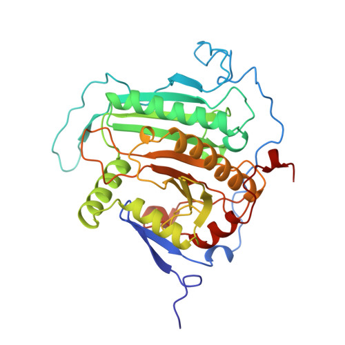
Structural analysis of the H166G site-directed mutant of galactose-1-phosphate uridylyltransferase complexed with either UDP-glucose or UDP-galactose: detailed description of the nucleotide sugar binding site.
Thoden, J.B., Ruzicka, F.J., Frey, P.A., Rayment, I., Holden, H.M.(1997) Biochemistry 36: 1212-1222
- PubMed: 9063869
- DOI: https://doi.org/10.1021/bi9626517
- Primary Citation of Related Structures:
1GUP, 1GUQ - PubMed Abstract:
Galactose-1-phosphate uridylyltransferase plays a key role in galactose metabolism by catalyzing the transfer of a uridine 5'-phosphoryl group from UDP-glucose to galactose 1-phosphate. The enzyme from Escherichia coli is composed of two identical subunits. The structures of the enzyme/UDP-glucose and UDP-galactose complexes, in which the catalytic nucleophile His 166 has been replaced with a glycine residue, have been determined and refined to 1.8 A resolution by single crystal X-ray diffraction analysis. Crystals employed in the investigation belonged to the space group P2(1) with unit cell dimensions of a = 68 A, b = 58 A, c = 189 A, and beta = 100 degrees and two dimers in the asymmetric unit. The models for these enzyme/substrate complexes have demonstrated that the active site of the uridylyltransferase is formed by amino acid residues contributed from both subunits in the dimer. Those amino acid residues critically involved in sugar binding include Asn 153 and Gly 159 from the first subunit and Lys 311, Phe 312, Val 314, Tyr 316, Glu 317, and Gln 323 from the second subunit. The uridylyltransferase is able to accommodate both UDP-galactose and UDP-glucose substrates by simple movements of the side chains of Glu 317 and Gln 323 and by a change in the backbone dihedral angles of Val 314. The removal of the imidazole group at position 166 results in little structural perturbation of the polypeptide chain backbone when compared to the previously determined structure for the wild-type enzyme. Instead, the cavity created by the mutation is partially compensated for by the presence of a potassium ion and its accompanying coordination sphere. As such, the mutant protein structures presented here represent valid models for understanding substrate recognition and binding in the native galactose-1-phosphate uridylyltransferase.
- Department of Biochemistry, University of Wisconsin-Madison, 53705, USA.
Organizational Affiliation:


