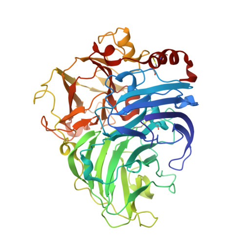
1GYC

CRYSTAL STRUCTURE DETERMINATION AT ROOM TEMPERATURE OF A LACCASE FROM TRAMETES VERSICOLOR IN ITS OXIDISED FORM CONTAINING A FULL COMPLEMENT OF COPPER IONS


Experimental Data Snapshot

  • Method: X-RAY DIFFRACTION
  • Resolution: 1.90 Å
  • R-Value Free: 0.212 
  • R-Value Work: 0.159 

Starting Model: experimental
View more details

wwPDB Validation   3D Report Full Report

Currently 1GYC does not have a validation slider image.


This is version 2.2 of the entry. See complete history


Literature

Crystal Structure of a Laccase from the Fungus Trametes Versicolor at 1.90-A Resolution Containing a Full Complement of Coppers.

Piontek, K.Antorini, M.Choinowski, T.

(2002) J Biol Chem 277: 37663

  • DOI: https://doi.org/10.1074/jbc.M204571200
  • Primary Citation of Related Structures:  
    1GYC

  • PubMed Abstract: 

    Laccase is a polyphenol oxidase, which belongs to the family of blue multicopper oxidases. These enzymes catalyze the one-electron oxidation of four reducing-substrate molecules concomitant with the four-electron reduction of molecular oxygen to water. Laccases oxidize a broad range of substrates, preferably phenolic compounds. In the presence of mediators, fungal laccases exhibit an enlarged substrate range and are then able to oxidize compounds with a redox potential exceeding their own. Until now, only one crystal structure of a laccase in an inactive, type-2 copper-depleted form has been reported. We present here the first crystal structure of an active laccase containing a full complement of coppers, the complete polypeptide chain together with seven carbohydrate moieties. Despite the presence of all coppers in the new structure, the folds of the two laccases are quite similar. The coordination of the type-3 coppers, however, is distinctly different. The geometry of the trinuclear copper cluster in the Trametes versicolor laccase is similar to that found in the ascorbate oxidase and that of mammalian ceruloplasmin structures, suggesting a common reaction mechanism for the copper oxidation and the O(2) reduction. In contrast to most blue copper proteins, the type-1 copper in the T. versicolor laccase has no axial ligand and is only 3-fold coordinated. Previously, a modest elevation of the redox potential was attributed to the lack of an axial ligand. Based on the present structural data and sequence comparisons, a mechanism is presented to explain how laccases could tune their redox potential by as much as 200 mV.


  • Organizational Affiliation

    Institute of Biochemistry, Swiss Federal Institute of Technology (ETH), ETH-Hönggerberg, Building HPM, Room D 8.1-2, CH-8093 Zürich, Switzerland. klaus.piontek@bc.biol.ethz.ch


Macromolecules
Find similar proteins by:  (by identity cutoff)  |  3D Structure
Entity ID: 1
MoleculeChains Sequence LengthOrganismDetailsImage
LACCASE 2499Trametes versicolorMutation(s): 1 
EC: 1.10.3.2
UniProt
Find proteins for Q12718 (Trametes versicolor)
Explore Q12718 
Go to UniProtKB:  Q12718
Entity Groups  
Sequence Clusters30% Identity50% Identity70% Identity90% Identity95% Identity100% Identity
UniProt GroupQ12718
Glycosylation
Glycosylation Sites: 6
Sequence Annotations
Expand
  • Reference Sequence
Oligosaccharides

Help

Entity ID: 2
MoleculeChains Length2D Diagram Glycosylation3D Interactions
2-acetamido-2-deoxy-beta-D-glucopyranose-(1-4)-2-acetamido-2-deoxy-beta-D-glucopyranose
B, C, D
2N-Glycosylation
Glycosylation Resources
GlyTouCan:  G42666HT
GlyCosmos:  G42666HT
GlyGen:  G42666HT
Small Molecules
Ligands 3 Unique
IDChains Name / Formula / InChI Key2D Diagram3D Interactions
NAG
Query on NAG

Download Ideal Coordinates CCD File 
I [auth A],
J [auth A],
K [auth A]
2-acetamido-2-deoxy-beta-D-glucopyranose
C8 H15 N O6
OVRNDRQMDRJTHS-FMDGEEDCSA-N
CU
Query on CU

Download Ideal Coordinates CCD File 
E [auth A],
F [auth A],
G [auth A],
H [auth A]
COPPER (II) ION
Cu
JPVYNHNXODAKFH-UHFFFAOYSA-N
IPA
Query on IPA

Download Ideal Coordinates CCD File 
L [auth A],
M [auth A]
ISOPROPYL ALCOHOL
C3 H8 O
KFZMGEQAYNKOFK-UHFFFAOYSA-N
Experimental Data & Validation

Experimental Data

  • Method: X-RAY DIFFRACTION
  • Resolution: 1.90 Å
  • R-Value Free: 0.212 
  • R-Value Work: 0.159 
  • Space Group: P 21 21 21
Unit Cell:
Length ( Å )Angle ( ˚ )
a = 83.681α = 90
b = 84.977β = 90
c = 91.81γ = 90
Software Package:
Software NamePurpose
REFMACrefinement
DENZOdata reduction
SCALEPACKdata scaling
AMoREphasing

Structure Validation

View Full Validation Report

Currently 1GYC does not have a validation slider image.


Entry History 

Deposition Data

Revision History  (Full details and data files)

  • Version 1.0: 2002-08-22
    Type: Initial release
  • Version 1.1: 2011-09-07
    Changes: Derived calculations, Non-polymer description, Other, Version format compliance
  • Version 1.2: 2019-07-24
    Changes: Data collection
  • Version 2.0: 2020-07-29
    Type: Remediation
    Reason: Carbohydrate remediation
    Changes: Atomic model, Data collection, Derived calculations, Structure summary
  • Version 2.1: 2023-12-13
    Changes: Data collection, Database references, Refinement description, Structure summary
  • Version 2.2: 2024-10-09
    Changes: Structure summary