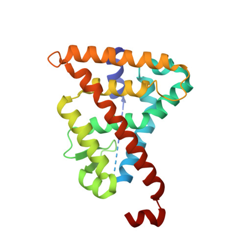
1H9U

The structure of the human retinoid-X-receptor beta ligand binding domain in complex with the specific synthetic agonist LG100268


Experimental Data Snapshot

  • Method: X-RAY DIFFRACTION
  • Resolution: 2.70 Å
  • R-Value Free: 0.306 
  • R-Value Work: 0.273 
  • R-Value Observed: 0.273 

Starting Model: experimental
View more details

wwPDB Validation   3D Report Full Report


This is version 1.3 of the entry. See complete history


Literature

The Structural Basis for the Specificity of Retinoid-X Receptor-Selective Agonists: New Insights Into the Role of Helix H12

Love, J.D.Gooch, J.T.Benko, S.Li, C.Nagy, L.Chatterjee, V.K.K.Evans, R.M.Schwabe, J.W.R.

(2002) J Biol Chem 277: 11385

  • DOI: https://doi.org/10.1074/jbc.M110869200
  • Primary Citation of Related Structures:  
    1H9U

  • PubMed Abstract: 

    Ligands that specifically target retinoid-X receptors (RXRs) are emerging as potentially powerful therapies for cancer, diabetes, and the lowering of circulatory cholesterol. To date, RXR has only been crystallized in the absence of ligand or with the promiscuous ligand 9-cis retinoic acid, which also activates retinoic acid receptors. Here we present the structure of hRXRbeta in complex with the RXR-specific agonist LG100268 (LG268). The structure clearly reveals why LG268 is specific for the RXR ligand binding pocket and will not activate retinoic acid receptors. Intriguingly, in the crystals, the C-terminal "activation" helix (AF-2/helix H12) is trapped in a novel position not seen in other nuclear receptor structures such that it does not cap the ligand binding cavity. Mammalian two-hybrid assays indicate that LG268 is unable to release co-repressors from RXR unless co-activators are also present. Together these findings suggest that RXR ligands may be inefficient at repositioning helix H12.


  • Organizational Affiliation

    Medical Research Council, Laboratory of Molecular Biology, Cambridge, United Kingdom.


Macromolecules
Find similar proteins by:  (by identity cutoff)  |  3D Structure
Entity ID: 1
MoleculeChains Sequence LengthOrganismDetailsImage
RETINOID X RECEPTOR, BETA
A, B, C, D
224Homo sapiensMutation(s): 0 
UniProt & NIH Common Fund Data Resources
Find proteins for P28702 (Homo sapiens)
Explore P28702 
Go to UniProtKB:  P28702
PHAROS:  P28702
GTEx:  ENSG00000204231 
Entity Groups  
Sequence Clusters30% Identity50% Identity70% Identity90% Identity95% Identity100% Identity
UniProt GroupP28702
Sequence Annotations
Expand
  • Reference Sequence
Small Molecules
Ligands 3 Unique
IDChains Name / Formula / InChI Key2D Diagram3D Interactions
LG2
Query on LG2

Download Ideal Coordinates CCD File 
E [auth A],
H [auth B],
K [auth C],
M [auth D]
6-[1-(3,5,5,8,8-PENTAMETHYL-5,6,7,8-TETRAHYDRONAPHTHALEN-2-YL)CYCLOPROPYL]PYRIDINE-3-CARBOXYLIC ACID
C24 H29 N O2
SLXTWXQUEZSSTJ-UHFFFAOYSA-N
NI
Query on NI

Download Ideal Coordinates CCD File 
F [auth A],
I [auth B],
L [auth C],
N [auth D]
NICKEL (II) ION
Ni
VEQPNABPJHWNSG-UHFFFAOYSA-N
CL
Query on CL

Download Ideal Coordinates CCD File 
G [auth A],
J [auth B]
CHLORIDE ION
Cl
VEXZGXHMUGYJMC-UHFFFAOYSA-M
Binding Affinity Annotations 
IDSourceBinding Affinity
LG2 BindingDB:  1H9U Ki: min: 3, max: 3515 (nM) from 7 assay(s)
Kd: min: 3, max: 3 (nM) from 2 assay(s)
IC50: min: 5, max: 8 (nM) from 2 assay(s)
EC50: min: 3, max: 40 (nM) from 6 assay(s)
Experimental Data & Validation

Experimental Data

  • Method: X-RAY DIFFRACTION
  • Resolution: 2.70 Å
  • R-Value Free: 0.306 
  • R-Value Work: 0.273 
  • R-Value Observed: 0.273 
  • Space Group: C 1 2 1
Unit Cell:
Length ( Å )Angle ( ˚ )
a = 123.886α = 90
b = 106.882β = 122.56
c = 100.587γ = 90
Software Package:
Software NamePurpose
CNSrefinement
MOSFLMdata reduction
SCALAdata scaling
CNSphasing

Structure Validation

View Full Validation Report


Entry History 

Deposition Data

Revision History  (Full details and data files)

  • Version 1.0: 2002-04-03
    Type: Initial release
  • Version 1.1: 2011-05-07
    Changes: Version format compliance
  • Version 1.2: 2011-07-13
    Changes: Version format compliance
  • Version 1.3: 2023-12-13
    Changes: Data collection, Database references, Derived calculations, Refinement description