1HKB | pdb_00001hkb

CRYSTAL STRUCTURE OF RECOMBINANT HUMAN BRAIN HEXOKINASE TYPE I COMPLEXED WITH GLUCOSE AND GLUCOSE-6-PHOSPHATE


Experimental Data Snapshot

  • Method: X-RAY DIFFRACTION
  • Resolution: 2.80 Å
  • R-Value Free: 
    0.270 (Depositor) 
  • R-Value Work: 
    0.200 (Depositor) 
  • R-Value Observed: 
    0.200 (Depositor) 

Starting Model: other
View more details

wwPDB Validation 3D Report Full Report

Validation slider image for 1HKB

Ligand Structure Quality Assessment 


This is version 1.5 of the entry. See complete history

Literature

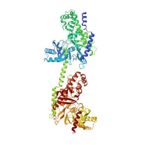
The mechanism of regulation of hexokinase: new insights from the crystal structure of recombinant human brain hexokinase complexed with glucose and glucose-6-phosphate.

Aleshin, A.E.Zeng, C.Bourenkov, G.P.Bartunik, H.D.Fromm, H.J.Honzatko, R.B.

(1998) Structure 6: 39-50

  • DOI: https://doi.org/10.1016/s0969-2126(98)00006-9
  • Primary Citation Related Structures: 
    1HKB

  • PubMed Abstract: 

    Hexokinase I is the pacemaker of glycolysis in brain tissue. The type I isozyme exhibits unique regulatory properties in that physiological levels of phosphate relieve potent inhibition by the product, glucose-6-phosphate (Gluc-6-P). The 100 kDa polypeptide chain of hexokinase I consists of a C-terminal (catalytic) domain and an N-terminal (regulatory) domain. Structures of ligated hexokinase I should provide a basis for understanding mechanisms of catalysis and regulation at an atomic level. The complex of human hexokinase I with glucose and Gluc-6-P (determined to 2.8 A resolution) is a dimer with twofold molecular symmetry. The N- and C-terminal domains of one monomer interact with the C- and N-terminal domains, respectively, of the symmetry-related monomer. The two domains of a monomer are connected by a single alpha helix and each have the fold of yeast hexokinase. Salt links between a possible cation-binding loop of the N-terminal domain and a loop of the C-terminal domain may be important to regulation. Each domain binds single glucose and Gluc-6-P molecules in proximity to each other. The 6-phosphoryl group of bound Gluc-6-P at the C-terminal domain occupies the putative binding site for ATP, whereas the 6-phosphoryl group at the N-terminal domain may overlap the binding site for phosphate. The binding synergism of glucose and Gluc-6-P probably arises out of the mutual stabilization of a common (glucose-bound) conformation of hexokinase I. Conformational changes in the N-terminal domain in response to glucose, phosphate, and/or Gluc-6-P may influence the binding of ATP to the C-terminal domain.


  • Organizational Affiliation
    • Department of Biochemistry and Biophysics, Iowa State University, Ames 50011, USA.

Macromolecule Content 

  • Total Structure Weight: 207.17 kDa 
  • Atom Count: 14,322 
  • Modeled Residue Count: 1,798 
  • Deposited Residue Count: 1,834 
  • Unique protein chains: 1

Macromolecules

Find similar proteins by:|  3D Structure
Entity ID: 1
MoleculeChains  Sequence LengthOrganismDetailsImage
D-GLUCOSE 6-PHOSPHOTRANSFERASE
A, B
917Homo sapiensMutation(s): 0 
EC: 2.7.1.1
UniProt & NIH Common Fund Data Resources
Find proteins for P19367 (Homo sapiens)
Explore P19367 
Go to UniProtKB:  P19367
PHAROS:  P19367
GTEx:  ENSG00000156515 
Entity Groups
Sequence Clusters30% Identity50% Identity70% Identity90% Identity95% Identity100% Identity
UniProt GroupP19367
Sequence Annotations
Expand
Reference Sequence

Small Molecules

Ligands 3 Unique
IDChains Name / Formula / InChI Key2D Diagram3D Interactions
G6P

Query on G6P


Download:Ideal Coordinates CCD File
D [auth A],
F [auth A],
J [auth B],
L [auth B]
6-O-phosphono-alpha-D-glucopyranose
C6 H13 O9 P
NBSCHQHZLSJFNQ-DVKNGEFBSA-N
BGC

Query on BGC


Download:Ideal Coordinates CCD File
C [auth A],
E [auth A],
I [auth B],
K [auth B]
beta-D-glucopyranose
C6 H12 O6
WQZGKKKJIJFFOK-VFUOTHLCSA-N
CA

Query on CA


Download:Ideal Coordinates CCD File
G [auth A],
H [auth A],
M [auth B],
N [auth B]
CALCIUM ION
Ca
BHPQYMZQTOCNFJ-UHFFFAOYSA-N

Experimental Data & Validation

Experimental Data

  • Method: X-RAY DIFFRACTION
  • Resolution: 2.80 Å
  • R-Value Free:  0.270 (Depositor) 
  • R-Value Work:  0.200 (Depositor) 
  • R-Value Observed: 0.200 (Depositor) 
Space Group: P 1 21 1
Unit Cell:
Length ( Å )Angle ( ˚ )
a = 83α = 90
b = 122β = 92
c = 123γ = 90
Software Package:
Software NamePurpose
X-PLORmodel building
X-PLORrefinement
DENZOdata reduction
SCALEPACKdata scaling
X-PLORphasing

Structure Validation

View Full Validation Report


Ligand Structure Quality Assessment 


Entry History 

Deposition Data

Revision History  (Full details and data files)

  • Version 1.0: 1998-06-03
    Type: Initial release
  • Version 1.1: 2008-03-03
    Changes: Version format compliance
  • Version 1.2: 2011-07-13
    Changes: Version format compliance
  • Version 1.3: 2020-07-29
    Type: Remediation
    Reason: Carbohydrate remediation
    Changes: Advisory, Data collection, Derived calculations, Other, Structure summary
  • Version 1.4: 2024-02-07
    Changes: Data collection, Database references, Structure summary
  • Version 1.5: 2024-04-03
    Changes: Refinement description