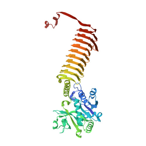
1HM8

CRYSTAL STRUCTURE OF S.PNEUMONIAE N-ACETYLGLUCOSAMINE-1-PHOSPHATE URIDYLTRANSFERASE, GLMU, BOUND TO ACETYL COENZYME A


Experimental Data Snapshot

  • Method: X-RAY DIFFRACTION
  • Resolution: 2.50 Å
  • R-Value Free: 0.243 
  • R-Value Work: 0.184 
  • R-Value Observed: 0.184 

wwPDB Validation   3D Report Full Report


Ligand Structure Quality Assessment 


This is version 1.3 of the entry. See complete history


Literature

Crystal structure of Streptococcus pneumoniae N-acetylglucosamine-1-phosphate uridyltransferase bound to acetyl-coenzyme A reveals a novel active site architecture.

Sulzenbacher, G.Gal, L.Peneff, C.Fassy, F.Bourne, Y.

(2001) J Biol Chem 276: 11844-11851

  • DOI: https://doi.org/10.1074/jbc.M011225200
  • Primary Citation of Related Structures:  
    1HM0, 1HM8, 1HM9

  • PubMed Abstract: 

    The bifunctional bacterial enzyme N-acetyl-glucosamine-1-phosphate uridyltransferase (GlmU) catalyzes the two-step formation of UDP-GlcNAc, a fundamental precursor in bacterial cell wall biosynthesis. With the emergence of new resistance mechanisms against beta-lactam and glycopeptide antibiotics, the biosynthetic pathway of UDP-GlcNAc represents an attractive target for drug design of new antibacterial agents. The crystal structures of Streptococcus pneumoniae GlmU in unbound form, in complex with acetyl-coenzyme A (AcCoA) and in complex with both AcCoA and the end product UDP-GlcNAc, have been determined and refined to 2.3, 2.5, and 1.75 A, respectively. The S. pneumoniae GlmU molecule is organized in two separate domains connected via a long alpha-helical linker and associates as a trimer, with the 50-A-long left-handed beta-helix (LbetaH) C-terminal domains packed against each other in a parallel fashion and the C-terminal region extended far away from the LbetaH core and exchanged with the beta-helix from a neighboring subunit in the trimer. AcCoA binding induces the formation of a long and narrow tunnel, enclosed between two adjacent LbetaH domains and the interchanged C-terminal region of the third subunit, giving rise to an original active site architecture at the junction of three subunits.


  • Organizational Affiliation

    AFMB-UMR6098, 31 Chemin Joseph Aiguier, 13402 Marseille Cedex 20, France.


Macromolecules
Find similar proteins by:  (by identity cutoff)  |  3D Structure
Entity ID: 1
MoleculeChains Sequence LengthOrganismDetailsImage
UDP-N-ACETYLGLUCOSAMINE-1-PHOSPHATE URIDYLTRANSFERASE
A, B
468Streptococcus pneumoniaeMutation(s): 0 
Gene Names: GLMU
EC: 2.7.7.23 (PDB Primary Data), 2.3.1.157 (UniProt)
UniProt
Find proteins for Q97R46 (Streptococcus pneumoniae serotype 4 (strain ATCC BAA-334 / TIGR4))
Explore Q97R46 
Go to UniProtKB:  Q97R46
Entity Groups  
Sequence Clusters30% Identity50% Identity70% Identity90% Identity95% Identity100% Identity
UniProt GroupQ97R46
Sequence Annotations
Expand
  • Reference Sequence
Experimental Data & Validation

Experimental Data

  • Method: X-RAY DIFFRACTION
  • Resolution: 2.50 Å
  • R-Value Free: 0.243 
  • R-Value Work: 0.184 
  • R-Value Observed: 0.184 
  • Space Group: H 3
Unit Cell:
Length ( Å )Angle ( ˚ )
a = 92.459α = 90
b = 92.459β = 90
c = 279.562γ = 120
Software Package:
Software NamePurpose
CNSrefinement
DENZOdata reduction
CCP4data scaling
CNSphasing

Structure Validation

View Full Validation Report


Ligand Structure Quality Assessment 


Entry History 

Deposition Data

Revision History  (Full details and data files)

  • Version 1.0: 2001-11-30
    Type: Initial release
  • Version 1.1: 2008-04-27
    Changes: Version format compliance
  • Version 1.2: 2011-07-13
    Changes: Derived calculations, Version format compliance
  • Version 1.3: 2024-02-07
    Changes: Data collection, Database references, Derived calculations