Structure of GSK3beta reveals a primed phosphorylation mechanism.
ter Haar, E., Coll, J.T., Austen, D.A., Hsiao, H.M., Swenson, L., Jain, J.(2001) Nat Struct Biol 8: 593-596
- PubMed: 11427888 Search on PubMed
- DOI: https://doi.org/10.1038/89624
- Primary Citation Related Structures:
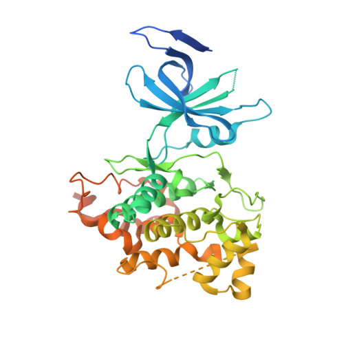
1I09 - PubMed Abstract:
GSK3beta was identified as the kinase that phosphorylates glycogen synthase but is now known to be involved in multiple signaling pathways. GSK3beta prefers prior phosphorylation of its substrates. We present the structure of unphosphorylated GSK3beta at 2.7 A. The orientation of the two domains and positioning of the activation loop of GSK3beta are similar to those observed in activated kinases. A phosphate ion held by Arg 96, Arg 180 and Lys 205 occupies the same position as the phosphate group of the phosphothreonine in activated p38gamma, CDK2 or ERK2. A loop from a neighboring molecule in the crystal occupies a portion of the substrate binding groove. The structure explains the unique primed phosphorylation mechanism of GSK3beta and how GSK3beta relies on a phosphoserine in the substrate for the alignment of the beta- and alpha-helical domains.
- Vertex Pharmaceuticals Incorporated, 130 Waverly Street, Cambridge, Massachusetts 02139-4211, USA. ernst_terhaar@vpharm.com
Organizational Affiliation: