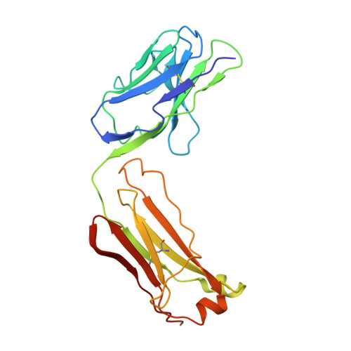
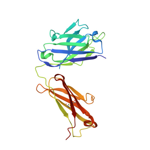
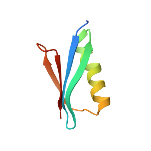
The third IgG-binding domain from streptococcal protein G. An analysis by X-ray crystallography of the structure alone and in a complex with Fab.
Derrick, J.P., Wigley, D.B.(1994) J Mol Biology 243: 906-918
- PubMed: 7966308
- DOI: https://doi.org/10.1006/jmbi.1994.1691
- Primary Citation of Related Structures:
1IGC, 1IGD - PubMed Abstract:
Protein G is a cell surface-associated protein from Streptococcus that binds to IgG with high affinity. We have determined the X-ray crystal structures of the third IgG-binding domain (domain III) alone to a resolution of 1.1 A (final R-factor of 19.3%), and in complex with an Fab fragment to 2.6 A (final R-factor of 16.8%). The structure of domain III is similar to the lower-resolution crystal structures of protein G domains determined previously by other investigators, but shows some minor differences when compared with the equivalent NMR structures. Domain III binds to the immunoglobulin by formation of an antiparallel interaction between the second beta-strand in domain III and the last beta-strand in the CH 1 domain. There is also a minor site of interaction between the C-terminal end of the alpha-helix in protein G and the first beta-strand in the CH 1 domain. Previous studies by NMR on the interactions between protein G and IgG have concluded that different portions of the protein G domain are involved in binding to the Fab and Fc portions. The results presented here support these findings and permit a detailed analysis of the recognition of Fab by protein G; formation of the complex buries a large water-accessible area, of a magnitude comparable with that found in antibody/antigen interactions. The majority of hydrogen bonds between the two proteins involve main-chain atoms from the CH 1 domain. The CH 1 domain residues that are in contact with protein G are shown to be highly conserved in alignments of mouse and human gamma chain amino acid sequences. We conclude that the binding site for protein G on Fab is relatively invariant across different species and gamma chain subclasses, providing an explanation for the widespread recognition of Fab fragments from mouse and human antibodies by protein G. The solution of the crystal structures of domain III alone and bound to Fab has demonstrated that there is no major structural change apparent in either protein on formation of the complex.
- Department of Biochemistry, University of Leicester, U.K.
Organizational Affiliation:


