1IIE | pdb_00001iie

HLA-DR ANTIGENS ASSOCIATED INVARIANT CHAIN


Experimental Data Snapshot

  • Method: SOLUTION NMR
  • Conformers Calculated: 80 
  • Conformers Submitted: 20 
  • Selection Criteria: LEAST RESTRAINT VIOLATION, GOOD GEOMETRY 

wwPDB Validation 3D Report Full Report

Validation slider image for 1IIE

This is version 1.5 of the entry. See complete history

Literature

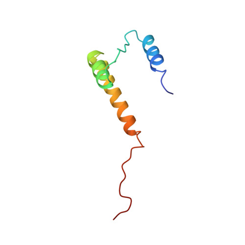
Structure of a trimeric domain of the MHC class II-associated chaperonin and targeting protein Ii.

Jasanoff, A.Wagner, G.Wiley, D.C.

(1998) EMBO J 17: 6812-6818

  • DOI: https://doi.org/10.1093/emboj/17.23.6812
  • Primary Citation Related Structures: 
    1IIE

  • PubMed Abstract: 

    The invariant chain (Ii) plays a critical role in MHC class II antigen processing by stabilizing peptide-free class II alphabeta heterodimers in a nonameric (alphabetaIi)3 complex soon after their synthesis and directing transport of the complex from the endoplasmic reticulum to compartments where peptide loading of class II takes place. Loading progresses following Ii proteolysis and via an intermediate complex of MHC class II with an Ii-derived peptide, CLIP. CLIP is substituted by exogenous peptidic fragments in an exchange reaction catalyzed by HLA-DM. The CLIP region of Ii, roughly residues 81-104, is one of two segments shown to interact with class II HLA-DR molecules. The other segment, Ii 118-216, is C-terminal to CLIP, mediates trimerization of the ectodomain of Ii and interferes with DM/class II binding. Here we report the three-dimensional structure of this trimeric domain, determined by nuclear magnetic resonance (NMR) studies of a 27 kDa trimer of human Ii 118-192. The cylindrical shape of the molecule and the mapping of conserved residues delimit surfaces which may be important for interactions between Ii and class II molecules.


  • Organizational Affiliation
    • Department of Molecular and Cellular Biology, Harvard Medical School, Cambridge, MA 02138, USA. jasanoff@crystal.harvard.edu

Macromolecule Content 

  • Total Structure Weight: 26.95 kDa 
  • Atom Count: 1,896 
  • Modeled Residue Count: 225 
  • Deposited Residue Count: 225 
  • Unique protein chains: 1

Macromolecules

Find similar proteins by:|  3D Structure
Entity ID: 1
MoleculeChains  Sequence LengthOrganismDetailsImage
PROTEIN (HLA-DR ANTIGENS ASSOCIATED INVARIANT CHAIN)
A, B, C
75Homo sapiensMutation(s): 0 
Gene Names: CD74 OR DHLAG
UniProt & NIH Common Fund Data Resources
Find proteins for P04233 (Homo sapiens)
Explore P04233 
Go to UniProtKB:  P04233
PHAROS:  P04233
GTEx:  ENSG00000019582 
Entity Groups
Sequence Clusters30% Identity50% Identity70% Identity90% Identity95% Identity100% Identity
UniProt GroupP04233
Sequence Annotations
Expand
Reference Sequence

Experimental Data & Validation

Experimental Data

  • Method: SOLUTION NMR
  • Conformers Calculated: 80 
  • Conformers Submitted: 20 
  • Selection Criteria: LEAST RESTRAINT VIOLATION, GOOD GEOMETRY 

Structure Validation

View Full Validation Report


Entry History 

Deposition Data

Revision History  (Full details and data files)

  • Version 1.0: 1999-02-16
    Type: Initial release
  • Version 1.1: 2008-04-26
    Changes: Version format compliance
  • Version 1.2: 2011-07-13
    Changes: Version format compliance
  • Version 1.3: 2019-11-06
    Changes: Data collection, Derived calculations
  • Version 1.4: 2021-10-27
    Changes: Data collection, Database references
  • Version 1.5: 2023-12-27
    Changes: Data collection