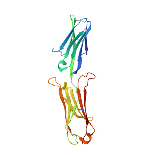
1IJ9

Highly Hydrated Human VCAM-1 Fragment


Experimental Data Snapshot

  • Method: X-RAY DIFFRACTION
  • Resolution: 3.00 Å
  • R-Value Free: 0.238 
  • R-Value Work: 0.208 
  • R-Value Observed: 0.208 

Starting Model: experimental
View more details

wwPDB Validation   3D Report Full Report


This is version 1.4 of the entry. See complete history


Literature

A new conformation of the integrin-binding fragment of human VCAM-1 crystallizes in a highly hydrated packing arrangement.

Taylor, P.Bilsland, M.Walkinshaw, M.D.

(2001) Acta Crystallogr D Biol Crystallogr 57: 1579-1583

  • DOI: https://doi.org/10.1107/s0907444901011209
  • Primary Citation of Related Structures:  
    1IJ9

  • PubMed Abstract: 

    An X-ray crystal structure of two N-terminal integrin-binding IgSF domains of human VCAM-1 is reported. This new crystal form shows an unusual and highly hydrated packing arrangement in which over 80% of the crystal is occupied by solvent. The relative orientations of the two domains adopt a new intermediate conformation. The tilt angle between the two domains is 19.4 degrees, compared with other related structures that have tilt angles ranging from 7.3 to 39.9 degrees. An analysis of the torsion angles shows that residues Ile88, Tyr89, Ser90, Pro92 and Glu96 play a major role in defining the interdomain conformations.


  • Organizational Affiliation

    Structural Biochemistry Group, Institute of Cell and Molecular Biology, The University of Edinburgh, Michael Swann Building, King's Buildings, Edinburgh EH9 3JR, Scotland.


Macromolecules
Find similar proteins by:  (by identity cutoff)  |  3D Structure
Entity ID: 1
MoleculeChains Sequence LengthOrganismDetailsImage
VASCULAR CELL ADHESION PROTEIN 1196Homo sapiensMutation(s): 0 
UniProt & NIH Common Fund Data Resources
Find proteins for P19320 (Homo sapiens)
Explore P19320 
Go to UniProtKB:  P19320
PHAROS:  P19320
GTEx:  ENSG00000162692 
Entity Groups  
Sequence Clusters30% Identity50% Identity70% Identity90% Identity95% Identity100% Identity
UniProt GroupP19320
Sequence Annotations
Expand
  • Reference Sequence
Experimental Data & Validation

Experimental Data

  • Method: X-RAY DIFFRACTION
  • Resolution: 3.00 Å
  • R-Value Free: 0.238 
  • R-Value Work: 0.208 
  • R-Value Observed: 0.208 
  • Space Group: P 31 2 1
Unit Cell:
Length ( Å )Angle ( ˚ )
a = 152.939α = 90
b = 152.939β = 90
c = 45.976γ = 120
Software Package:
Software NamePurpose
DENZOdata reduction
SCALEPACKdata scaling
AMoREphasing
X-PLORrefinement

Structure Validation

View Full Validation Report


Entry History 

Deposition Data

Revision History  (Full details and data files)

  • Version 1.0: 2001-05-02
    Type: Initial release
  • Version 1.1: 2008-04-27
    Changes: Version format compliance
  • Version 1.2: 2011-07-13
    Changes: Version format compliance
  • Version 1.3: 2023-08-16
    Changes: Data collection, Database references, Refinement description
  • Version 1.4: 2024-10-30
    Changes: Structure summary