1IJY | pdb_00001ijy

CRYSTAL STRUCTURE OF THE CYSTEINE-RICH DOMAIN OF MOUSE FRIZZLED 8 (MFZ8)


Experimental Data Snapshot

  • Method: X-RAY DIFFRACTION
  • Resolution: 1.35 Å
  • R-Value Free: 
    0.247 (Depositor), 0.248 (DCC) 
  • R-Value Work: 
    0.225 (Depositor), 0.226 (DCC) 

Starting Model: experimental
View more details

wwPDB Validation 3D Report Full Report

Validation slider image for 1IJY

This is version 1.6 of the entry. See complete history

Literature

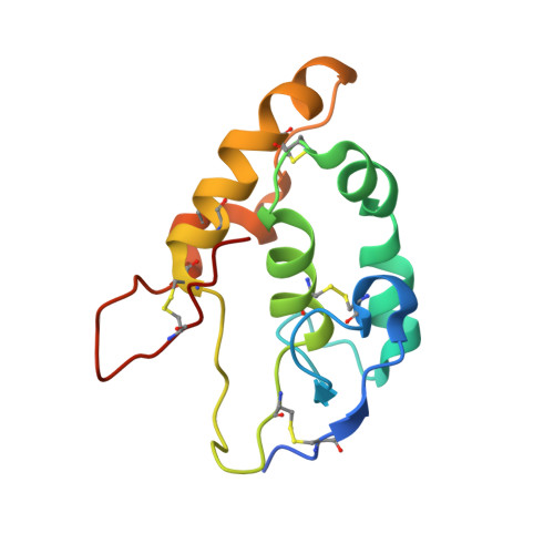
Insights into Wnt binding and signalling from the structures of two Frizzled cysteine-rich domains.

Dann III, C.E.Hsieh, J.C.Rattner, A.Sharma, D.Nathans, J.Leahy, D.J.

(2001) Nature 412: 86-90

  • DOI: https://doi.org/10.1038/35083601
  • Primary Citation Related Structures: 
    1IJX, 1IJY

  • PubMed Abstract: 

    Members of the Frizzled family of seven-pass transmembrane proteins serve as receptors for Wnt signalling proteins. Wnt proteins have important roles in the differentiation and patterning of diverse tissues during animal development, and inappropriate activation of Wnt signalling pathways is a key feature of many cancers. An extracellular cysteine-rich domain (CRD) at the amino terminus of Frizzled proteins binds Wnt proteins, as do homologous domains in soluble proteins-termed secreted Frizzled-related proteins-that function as antagonists of Wnt signalling. Recently, an LDL-receptor-related protein has been shown to function as a co-receptor for Wnt proteins and to bind to a Frizzled CRD in a Wnt-dependent manner. To investigate the molecular nature of the Wnt signalling complex, we determined the crystal structures of the CRDs from mouse Frizzled 8 and secreted Frizzled-related protein 3. Here we show a previously unknown protein fold, and the design and interpretation of CRD mutations that identify a Wnt-binding site. CRDs exhibit a conserved dimer interface that may be a feature of Wnt signalling. This work provides a framework for studies of homologous CRDs in proteins including muscle-specific kinase and Smoothened, a component of the Hedgehog signalling pathway.


  • Organizational Affiliation
    • Department of Biophysics and Biophysical Chemistry, Johns Hopkins University School of Medicine, Baltimore, MD 21205, USA.

Macromolecule Content 

  • Total Structure Weight: 29.89 kDa 
  • Atom Count: 2,257 
  • Modeled Residue Count: 244 
  • Deposited Residue Count: 260 
  • Unique protein chains: 1

Macromolecules

Find similar proteins by:|  3D Structure
Entity ID: 1
MoleculeChains  Sequence LengthOrganismDetailsImage
FRIZZLED HOMOLOG 8
A, B
130Mus musculusMutation(s): 2 
Gene Names: FZD8
UniProt & NIH Common Fund Data Resources
Find proteins for Q61091 (Mus musculus)
Explore Q61091 
Go to UniProtKB:  Q61091
IMPC:  MGI:108460
Entity Groups
Sequence Clusters30% Identity50% Identity70% Identity90% Identity95% Identity100% Identity
UniProt GroupQ61091
Sequence Annotations
Expand
Reference Sequence

Experimental Data & Validation

Experimental Data

  • Method: X-RAY DIFFRACTION
  • Resolution: 1.35 Å
  • R-Value Free:  0.247 (Depositor), 0.248 (DCC) 
  • R-Value Work:  0.225 (Depositor), 0.226 (DCC) 
Space Group: P 1 2 1
Unit Cell:
Length ( Å )Angle ( ˚ )
a = 55.69α = 90
b = 36.37β = 98.82
c = 57.94γ = 90
Software Package:
Software NamePurpose
DENZOdata reduction
SCALEPACKdata scaling
CNSrefinement
CNSphasing

Structure Validation

View Full Validation Report


Entry History 

Deposition Data

Revision History  (Full details and data files)

  • Version 1.0: 2001-07-11
    Type: Initial release
  • Version 1.1: 2008-04-27
    Changes: Version format compliance
  • Version 1.2: 2011-07-13
    Changes: Version format compliance
  • Version 1.3: 2014-05-28
    Changes: Structure summary
  • Version 1.4: 2021-10-27
    Changes: Database references
  • Version 1.5: 2023-08-16
    Changes: Data collection, Refinement description
  • Version 1.6: 2024-10-30
    Changes: Structure summary