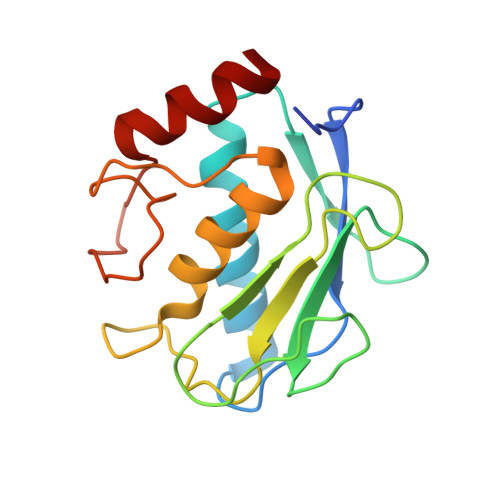
The 1.8-A crystal structure of a matrix metalloproteinase 8-barbiturate inhibitor complex reveals a previously unobserved mechanism for collagenase substrate recognition.
Brandstetter, H., Grams, F., Glitz, D., Lang, A., Huber, R., Bode, W., Krell, H.W., Engh, R.A.(2001) J Biological Chem 276: 17405-17412
- PubMed: 11278347 Search on PubMed
- DOI: https://doi.org/10.1074/jbc.M007475200
- Primary Citation Related Structures:
1JJ9 - PubMed Abstract:
The individual zinc endoproteinases of the tissue degrading matrix metalloproteinase (MMP) family share a common catalytic architecture but are differentiated with respect to substrate specificity, localization, and activation. Variation in domain structure and more subtle structural differences control their characteristic specificity profiles for substrates from among four distinct classes (Nagase, H., and Woessner, J. F. J. (1999) J. Biol. Chem. 274, 21491-21494). Exploitation of these differences may be decisive for the design of anticancer or other drugs, which should be highly selective for their particular MMP targets. Based on the 1.8-A crystal structure of human neutrophil collagenase (MMP-8) in complex with an active site-directed inhibitor (RO200-1770), we identify and describe new structural determinants for substrate and inhibitor recognition in addition to the primary substrate recognition sites. RO200-1770 induces a major rearrangement at a position relevant to substrate recognition near the MMP-8 active site (Ala206-Asn218). In stromelysin (MMP-3), competing stabilizing interactions at the analogous segment hinder a similar rearrangement, consistent with kinetic profiling of several MMPs. Despite the apparent dissimilarity of the inhibitors, the central 2-hydroxypyrimidine-4,6-dione (barbiturate) ring of the inhibitor RO200-1770 mimics the interactions of the hydroxamate-derived inhibitor batimastat (Grams, F., Reinemer, P., Powers, J. C., Kleine, T., Pieper, M., Tschesche, H., Huber, R., and Bode, W. (1995) Eur. J. Biochem. 228, 830-841) for binding to MMP-8. The two additional phenyl and piperidyl ring substituents of the inhibitor bind into the S1' and S2' pockets of MMP-8, respectively. The crystal lattice contains a hydrogen bond between the O(gamma) group of Ser209 and N(delta)1 of His207 of a symmetry related molecule; this interaction suggests a model for recognition of hydroxyprolines present in physiological substrates. We also identify a collagenase-characteristic cis-peptide bond, Asn188-Tyr189, on a loop essential for collagenolytic activity. The sequence conservation pattern at this position marks this cis-peptide bond as a determinant for triple-helical collagen recognition and processing.
- Department of Structural Research, Max Planck Institute for Biochemistry, D-82152 Martinsried, Germany. hbs@biochem.mpg.de
Organizational Affiliation: