1JLW | pdb_00001jlw

Anopheles dirus species B glutathione S-transferases 1-4


Experimental Data Snapshot

  • Method: X-RAY DIFFRACTION
  • Resolution: 2.45 Å
  • R-Value Free: 
    0.271 (Depositor), 0.269 (DCC) 
  • R-Value Work: 
    0.223 (Depositor), 0.223 (DCC) 
  • R-Value Observed: 
    0.223 (Depositor) 

Starting Model: other
View more details

wwPDB Validation 3D Report Full Report

Validation slider image for 1JLW

This is version 1.4 of the entry. See complete history

Literature

The crystal structures of glutathione S-transferases isozymes 1-3 and 1-4 from Anopheles dirus species B.

Oakley, A.J.Harnnoi, T.Udomsinprasert, R.Jirajaroenrat, K.Ketterman, A.J.Wilce, M.C.

(2001) Protein Sci 10: 2176-2185

  • DOI: https://doi.org/10.1110/ps.ps.21201
  • Primary Citation Related Structures: 
    1JLV, 1JLW

  • PubMed Abstract: 

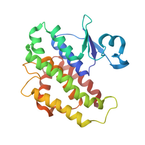
    Glutathione S-transferases (GSTs) are dimeric proteins that play an important role in cellular detoxification. Four GSTs from the mosquito Anopheles dirus species B (Ad), an important malaria vector in South East Asia, are produced by alternate splicing of a single transcription product and were previously shown to have detoxifying activity towards pesticides such as DDT. We have determined the crystal structures for two of these alternatively spliced proteins, AdGST1-3 (complexed with glutathione) and AdGST1-4 (apo form), at 1.75 and 2.45 A resolution, respectively. These GST isozymes show differences from the related GST from the Australian sheep blowfly Lucilia cuprina; in particular, the presence of a C-terminal helix forming part of the active site. This helix causes the active site of the Anopheles GSTs to be enclosed. The glutathione-binding helix alpha2 and flanking residues are disordered in the AdGST1-4 (apo) structure, yet ordered in the AdGST1-3 (GSH-bound) structure, suggesting that insect GSTs operate with an induced fit mechanism similar to that found in the plant phi- and human pi-class GSTs. Despite the high overall sequence identities, the active site residues of AdGST1-4 and AdGST1-3 have different conformations.


  • Organizational Affiliation
    • Department of Pharmacology/Crystallography Centre, University of Western Australia, Crawley 6009, Australia.

Macromolecule Content 

  • Total Structure Weight: 50 kDa 
  • Atom Count: 3,532 
  • Modeled Residue Count: 434 
  • Deposited Residue Count: 438 
  • Unique protein chains: 1

Macromolecules

Find similar proteins by:|  3D Structure
Entity ID: 1
MoleculeChains  Sequence LengthOrganismDetailsImage
glutathione transferase GST1-4
A, B
219Anopheles cracensMutation(s): 0 
EC: 2.5.1.18
UniProt
Find proteins for Q7KIF1 (Anopheles cracens)
Explore Q7KIF1 
Go to UniProtKB:  Q7KIF1
Entity Groups
Sequence Clusters30% Identity50% Identity70% Identity90% Identity95% Identity100% Identity
UniProt GroupQ7KIF1
Sequence Annotations
Expand
Reference Sequence

Experimental Data & Validation

Experimental Data

  • Method: X-RAY DIFFRACTION
  • Resolution: 2.45 Å
  • R-Value Free:  0.271 (Depositor), 0.269 (DCC) 
  • R-Value Work:  0.223 (Depositor), 0.223 (DCC) 
  • R-Value Observed: 0.223 (Depositor) 
Space Group: P 32 2 1
Unit Cell:
Length ( Å )Angle ( ˚ )
a = 49.44α = 90
b = 49.44β = 90
c = 271.81γ = 120
Software Package:
Software NamePurpose
DENZOdata reduction
SCALEPACKdata scaling
MOLREPphasing
CNSrefinement

Structure Validation

View Full Validation Report


Entry History 

Deposition Data

Revision History  (Full details and data files)

  • Version 1.0: 2002-07-16
    Type: Initial release
  • Version 1.1: 2008-04-27
    Changes: Version format compliance
  • Version 1.2: 2011-07-13
    Changes: Version format compliance
  • Version 1.3: 2024-02-07
    Changes: Data collection, Database references
  • Version 1.4: 2024-04-03
    Changes: Refinement description