1JQ8

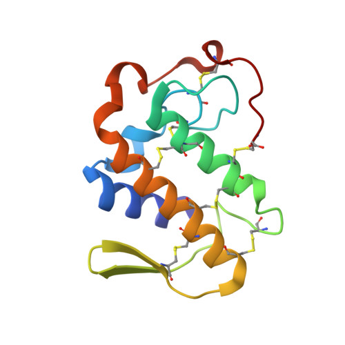
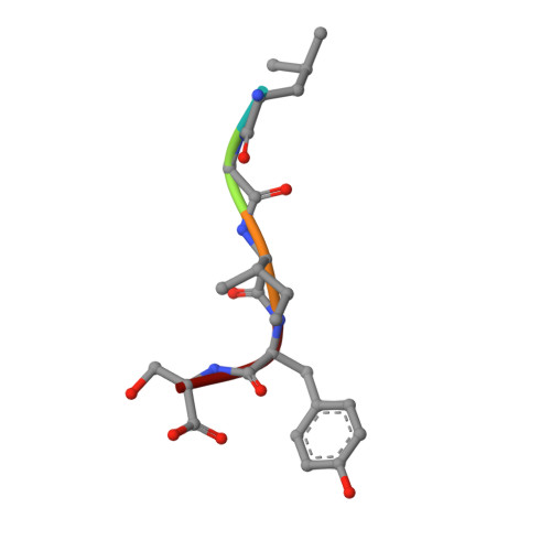
Design of specific inhibitors of phospholipase A2: Crystal structure of a complex formed between phospholipase A2 from Daboia russelli pulchella and a designed pentapeptide Leu-Ala-Ile-Tyr-Ser at 2.0 resolution


Experimental Data Snapshot

  • Method: X-RAY DIFFRACTION
  • Resolution: 2.00 Å
  • R-Value Free: 0.198 
  • R-Value Work: 0.187 
  • R-Value Observed: 0.187 

Starting Model: experimental
View more details

wwPDB Validation   3D Report Full Report


This is version 1.6 of the entry. See complete history

Re-refinement Note

A newer entry is available that reflects an alternative modeling of the original data: 5VET


Literature

Design of specific peptide inhibitors of phospholipase A2: structure of a complex formed between Russell's viper phospholipase A2 and a designed peptide Leu-Ala-Ile-Tyr-Ser (LAIYS).

Chandra, V.Jasti, J.Kaur, P.Dey, S.Srinivasan, A.Betzel, C.h.Singh, T.P.

(2002) Acta Crystallogr D Biol Crystallogr 58: 1813-1819

  • DOI: https://doi.org/10.1107/s0907444902013720
  • Primary Citation of Related Structures:  
    1JQ8

  • PubMed Abstract: 

    Phospholipase A(2) (EC 3.1.1.4) is a key enzyme of the cascade mechanism involved in the production of proinflammatory compounds known as eicosanoids. The binding of phospholipase A(2) to membrane surfaces and the hydrolysis of phospholipids are thought to involve the formation of a hydrophobic channel into which a single substrate molecule diffuses before cleavage. In order to regulate the production of proinflammatory compounds, a specific peptide inhibitor of PLA(2), Leu-Ala-Ile-Tyr-Ser, has been designed. Phospholipase A(2) from Daboia russelli pulchella (DPLA(2)) and peptide Leu-Ala-Ile-Tyr-Ser (LAIYS) have been co-crystallized. The structure of the complex has been determined and refined to 2.0 A resolution. The structure contains two crystallographically independent molecules of DPLA(2), with one molecule of peptide specifically bound to one of them. The overall conformations of the two molecules are essentially similar except in three regions; namely, the calcium-binding loop including Trp31 (residues 25-34), the beta-wing consisting of two antiparallel beta-strands (residues 74-85) and the C-terminal region (residues 119-133). Of these, the most striking difference pertains to the orientation of Trp31 in the two molecules. The conformation of Trp31 in molecule A was suitable to allow the binding of peptide LAIYS, while that in molecule B prevented the entry of the ligand into the hydrophobic channel. The structure of the complex clearly showed that the OH group of Tyr of the inhibitor formed hydrogen bonds with both His48 N(delta1) and Asp49 O(delta1), while O(gamma)H of Ser was involved in a hydrogen bond with Trp31. Other peptide backbone atoms interact with protein through water molecules, while Leu, Ala and Ile form strong hydrophobic interactions with the residues of the hydrophobic channel.


  • Organizational Affiliation

    Department of Biophysics, All India Institute of Medical Sciences, New Delhi 110029, India.


Macromolecules
Find similar proteins by:  (by identity cutoff)  |  3D Structure
Entity ID: 1
MoleculeChains Sequence LengthOrganismDetailsImage
Phospholipase A2
A, B
121Daboia russelii pulchellaMutation(s): 0 
EC: 3.1.1.4
UniProt
Find proteins for P59071 (Daboia russelii)
Explore P59071 
Go to UniProtKB:  P59071
Entity Groups  
Sequence Clusters30% Identity50% Identity70% Identity90% Identity95% Identity100% Identity
UniProt GroupP59071
Sequence Annotations
Expand
  • Reference Sequence

Find similar proteins by:  Sequence   |   3D Structure  

Entity ID: 2
MoleculeChains Sequence LengthOrganismDetailsImage
Peptide inhibitorC [auth P]5N/AMutation(s): 0 
Sequence Annotations
Expand
  • Reference Sequence
Experimental Data & Validation

Experimental Data

  • Method: X-RAY DIFFRACTION
  • Resolution: 2.00 Å
  • R-Value Free: 0.198 
  • R-Value Work: 0.187 
  • R-Value Observed: 0.187 
  • Space Group: C 2 2 21
Unit Cell:
Length ( Å )Angle ( ˚ )
a = 76.825α = 90
b = 90.375β = 90
c = 77.59γ = 90
Software Package:
Software NamePurpose
MAR345data collection
SCALEPACKdata scaling
AMoREphasing
CNSrefinement

Structure Validation

View Full Validation Report


Entry History 

Deposition Data

Revision History  (Full details and data files)

  • Version 1.0: 2002-11-06
    Type: Initial release
  • Version 1.1: 2008-04-27
    Changes: Version format compliance
  • Version 1.2: 2011-07-13
    Changes: Atomic model, Database references, Derived calculations, Non-polymer description, Structure summary, Version format compliance
  • Version 1.3: 2011-11-16
    Changes: Atomic model
  • Version 1.4: 2017-10-04
    Changes: Refinement description
  • Version 1.5: 2019-10-23
    Changes: Data collection
  • Version 1.6: 2023-10-25
    Changes: Data collection, Database references, Derived calculations, Refinement description