1JU3 | pdb_00001ju3

BACTERIAL COCAINE ESTERASE COMPLEX WITH TRANSITION STATE ANALOG


Experimental Data Snapshot

  • Method: X-RAY DIFFRACTION
  • Resolution: 1.58 Å
  • R-Value Free: 
    0.214 (Depositor), 0.211 (DCC) 
  • R-Value Work: 
    0.199 (Depositor), 0.195 (DCC) 
  • R-Value Observed: 
    0.199 (Depositor) 

Starting Model: experimental
View more details

wwPDB Validation 3D Report Full Report

Validation slider image for 1JU3

This is version 1.5 of the entry. See complete history

Literature

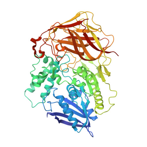
Crystal structure of a bacterial cocaine esterase.

Larsen, N.A.Turner, J.M.Stevens, J.Rosser, S.J.Basran, A.Lerner, R.A.Bruce, N.C.Wilson, I.A.

(2002) Nat Struct Biol 9: 17-21

  • DOI: https://doi.org/10.1038/nsb742
  • Primary Citation Related Structures: 
    1JU3, 1JU4

  • PubMed Abstract: 

    Here we report the first structure of a cocaine-degrading enzyme. The bacterial esterase, cocE, hydrolyzes pharmacologically active (-)-cocaine to a non-psychoactive metabolite with a rate faster than any other reported cocaine esterase (kcat = 7.8 s-1 and KM = 640 nM). Because of the high catalytic proficiency of cocE, it is an attractive candidate for novel protein-based therapies for cocaine overdose. The crystal structure of cocE, solved by multiple anomalous dispersion (MAD) methods, reveals that cocE is a serine esterase composed of three domains: (i) a canonical alpha/beta hydrolase fold (ii) an alpha-helical domain that caps the active site and (iii) a jelly-roll-like beta-domain that interacts extensively with the other two domains. The active site was identified within the interface of all three domains by analysis of the crystal structures of transition state analog adduct and product complexes, which were refined at 1.58 A and 1.63 A resolution, respectively. These structural studies suggest that substrate recognition arises partly from interactions between the benzoyl moiety of cocaine and a highly evolved specificity pocket.


  • Organizational Affiliation
    • Department of Molecular Biology, The Scripps Research Institute, 10550 North Torrey Pines Rd., La Jolla, California 92037, USA.

Macromolecule Content 

  • Total Structure Weight: 63.47 kDa 
  • Atom Count: 4,904 
  • Modeled Residue Count: 570 
  • Deposited Residue Count: 583 
  • Unique protein chains: 1

Macromolecules

Find similar proteins by:|  3D Structure
Entity ID: 1
MoleculeChains  Sequence LengthOrganismDetailsImage
cocaine esterase583Rhodococcus sp. MB1Mutation(s): 0 
EC: 3.1.1.84
UniProt
Find proteins for Q9L9D7 (Rhodococcus sp. (strain MB1 Bresler))
Explore Q9L9D7 
Go to UniProtKB:  Q9L9D7
Entity Groups
Sequence Clusters30% Identity50% Identity70% Identity90% Identity95% Identity100% Identity
UniProt GroupQ9L9D7
Sequence Annotations
Expand
Reference Sequence

Small Molecules

Ligands 1 Unique
IDChains Name / Formula / InChI Key2D Diagram3D Interactions
PBC

Query on PBC


Download:Ideal Coordinates CCD File
B [auth A]PHENYL BORONIC ACID
C6 H7 B O2
HXITXNWTGFUOAU-UHFFFAOYSA-N

Experimental Data & Validation

Experimental Data

  • Method: X-RAY DIFFRACTION
  • Resolution: 1.58 Å
  • R-Value Free:  0.214 (Depositor), 0.211 (DCC) 
  • R-Value Work:  0.199 (Depositor), 0.195 (DCC) 
  • R-Value Observed: 0.199 (Depositor) 
Space Group: P 65 2 2
Unit Cell:
Length ( Å )Angle ( ˚ )
a = 106.08α = 90
b = 106.08β = 90
c = 221.52γ = 120
Software Package:
Software NamePurpose
CNSrefinement
DENZOdata reduction
SCALEPACKdata scaling
CNSphasing

Structure Validation

View Full Validation Report


Entry History 

Deposition Data

Revision History  (Full details and data files)

  • Version 1.0: 2001-12-21
    Type: Initial release
  • Version 1.1: 2007-10-16
    Changes: Version format compliance
  • Version 1.2: 2011-07-13
    Changes: Version format compliance
  • Version 1.3: 2018-01-31
    Changes: Experimental preparation
  • Version 1.4: 2023-08-16
    Changes: Data collection, Database references, Derived calculations, Refinement description
  • Version 1.5: 2024-10-16
    Changes: Structure summary