1JXQ

Structure of cleaved, CARD domain deleted Caspase-9


Experimental Data Snapshot

  • Method: X-RAY DIFFRACTION
  • Resolution: 2.80 Å
  • R-Value Free: 0.275 
  • R-Value Work: 0.233 

Starting Model: experimental
View more details

wwPDB Validation   3D Report Full Report


This is version 1.4 of the entry. See complete history


Literature

Dimer formation drives the activation of the cell death protease caspase 9.

Renatus, M.Stennicke, H.R.Scott, F.L.Liddington, R.C.Salvesen, G.S.

(2001) Proc Natl Acad Sci U S A 98: 14250-14255

  • DOI: https://doi.org/10.1073/pnas.231465798
  • Primary Citation of Related Structures:  
    1JXQ

  • PubMed Abstract: 

    A critical step in the induction of apoptosis is the activation of the apoptotic initiator caspase 9. We show that at its normal physiological concentration, caspase 9 is primarily an inactive monomer (zymogen), and that activity is associated with a dimeric species. At the high concentrations used for crystal formation, caspase 9 is dimeric, and the structure reveals two very different active-site conformations within each dimer. One site closely resembles the catalytically competent sites of other caspases, whereas in the second, expulsion of the "activation loop" disrupts the catalytic machinery. We propose that the inactive domain resembles monomeric caspase 9. Activation is induced by dimerization, with interactions at the dimer interface promoting reorientation of the activation loop. These observations support a model in which recruitment by Apaf-1 creates high local concentrations of caspase 9 to provide a pathway for dimer-induced activation.


  • Organizational Affiliation

    Program in Apoptosis and Cell Death Research, The Burnham Institute, La Jolla, CA 92037, USA.


Macromolecules
Find similar proteins by:  (by identity cutoff)  |  3D Structure
Entity ID: 1
MoleculeChains Sequence LengthOrganismDetailsImage
Caspase-9
A, B, C, D
284Homo sapiensMutation(s): 0 
EC: 3.4.22 (PDB Primary Data), 3.4.22.62 (UniProt)
UniProt & NIH Common Fund Data Resources
Find proteins for P55211 (Homo sapiens)
Explore P55211 
Go to UniProtKB:  P55211
PHAROS:  P55211
GTEx:  ENSG00000132906 
Entity Groups  
Sequence Clusters30% Identity50% Identity70% Identity90% Identity95% Identity100% Identity
UniProt GroupP55211
Sequence Annotations
Expand
  • Reference Sequence

Find similar proteins by:  Sequence   |   3D Structure  

Entity ID: 2
MoleculeChains Sequence LengthOrganismDetailsImage
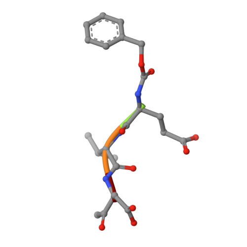
benzoxycarbonyl-Val-Ala-Asp-fluoromethyl ketone Inhibitor
E, F
5N/AMutation(s): 0 
Sequence Annotations
Expand
  • Reference Sequence
Experimental Data & Validation

Experimental Data

  • Method: X-RAY DIFFRACTION
  • Resolution: 2.80 Å
  • R-Value Free: 0.275 
  • R-Value Work: 0.233 
  • Space Group: C 1 2 1
Unit Cell:
Length ( Å )Angle ( ˚ )
a = 144.3α = 90
b = 81.8β = 111.7
c = 125.4γ = 90
Software Package:
Software NamePurpose
AMoREphasing
CNSrefinement
DENZOdata reduction
SCALEPACKdata scaling

Structure Validation

View Full Validation Report


Entry History 

Deposition Data

Revision History  (Full details and data files)

  • Version 1.0: 2001-12-12
    Type: Initial release
  • Version 1.1: 2008-04-27
    Changes: Version format compliance
  • Version 1.2: 2011-07-13
    Changes: Atomic model, Database references, Derived calculations, Non-polymer description, Structure summary, Version format compliance
  • Version 1.3: 2023-08-16
    Changes: Data collection, Database references, Derived calculations, Refinement description
  • Version 1.4: 2024-11-13
    Changes: Structure summary