1KI0

The X-ray Structure of Human Angiostatin


Experimental Data Snapshot

  • Method: X-RAY DIFFRACTION
  • Resolution: 1.75 Å
  • R-Value Free: 0.263 
  • R-Value Work: 0.196 

wwPDB Validation   3D Report Full Report


This is version 1.3 of the entry. See complete history


Literature

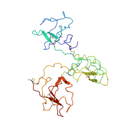
The X-ray crystallographic structure of the angiogenesis inhibitor angiostatin.

Abad, M.C.Arni, R.K.Grella, D.K.Castellino, F.J.Tulinsky, A.Geiger, J.H.

(2002) J Mol Biol 318: 1009-1017

  • DOI: https://doi.org/10.1016/S0022-2836(02)00211-5
  • Primary Citation of Related Structures:  
    1KI0

  • PubMed Abstract: 

    Angiogenesis inhibitors have gained much public attention recently as anti-cancer agents and several are currently in clinical trials, including angiostatin (Phase I, Thomas Jefferson University Hospital, Philadelphia, PA). We report here the bowl-shaped structure of angiostatin kringles 1-3, the first multi-kringle structure to be determined. All three kringle lysine-binding sites contain a bound bicine molecule of crystallization while the former of kringle 2 and kringle 3 are cofacial. Moreover, the separation of the kringle 2 and kringle 3 lysiner binding sites is sufficient to accommodate the alpha-helix of the 30 residue peptide VEK-30 found in the kringle 2/VEK-30 complex. Together the three kringles produce a central cavity suggestive of a unique domain where they may function in concert.


  • Organizational Affiliation

    Department of Chemistry, Michigan State University, East Lansing 48824, USA.


Macromolecules
Find similar proteins by:  (by identity cutoff)  |  3D Structure
Entity ID: 1
MoleculeChains Sequence LengthOrganismDetailsImage
ANGIOSTATIN253Homo sapiensMutation(s): 1 
EC: 3.4.21.7
UniProt & NIH Common Fund Data Resources
Find proteins for P00747 (Homo sapiens)
Explore P00747 
Go to UniProtKB:  P00747
PHAROS:  P00747
GTEx:  ENSG00000122194 
Entity Groups  
Sequence Clusters30% Identity50% Identity70% Identity90% Identity95% Identity100% Identity
UniProt GroupP00747
Sequence Annotations
Expand
  • Reference Sequence
Experimental Data & Validation

Experimental Data

  • Method: X-RAY DIFFRACTION
  • Resolution: 1.75 Å
  • R-Value Free: 0.263 
  • R-Value Work: 0.196 
  • Space Group: P 41 21 2
Unit Cell:
Length ( Å )Angle ( ˚ )
a = 56.943α = 90
b = 56.943β = 90
c = 192.966γ = 90
Software Package:
Software NamePurpose
DENZOdata reduction
SCALEPACKdata scaling
AMoREphasing
CNSrefinement

Structure Validation

View Full Validation Report


Entry History 

Deposition Data

Revision History  (Full details and data files)

  • Version 1.0: 2002-05-29
    Type: Initial release
  • Version 1.1: 2008-04-27
    Changes: Version format compliance
  • Version 1.2: 2011-07-13
    Changes: Version format compliance
  • Version 1.3: 2021-10-27
    Changes: Database references, Derived calculations