Structural and Functional Evidence for Ligand-Independent Transcriptional Activation by the Estrogen-Related Receptor 3
Greschik, H., Wurtz, J.-M., Sanglier, S., Bourguet, W., van Dorsselaer, A., Moras, D., Renaud, J.-P.(2002) Mol Cell 9: 303-313
- PubMed: 11864604 Search on PubMed
- DOI: https://doi.org/10.1016/s1097-2765(02)00444-6
- Primary Citation Related Structures:
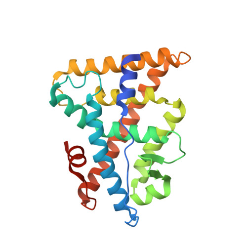
1KV6 - PubMed Abstract:
The crystal structure of the ligand binding domain (LBD) of the estrogen-related receptor 3 (ERR3) complexed with a steroid receptor coactivator-1 (SRC-1) peptide reveals a transcriptionally active conformation in absence of any ligand. The structure explains why estradiol does not bind ERRs with significant affinity. Docking of the previously reported ERR antagonists, diethylstilbestrol and 4-hydroxytamoxifen, requires structural rearrangements enlarging the ligand binding pocket that can only be accommodated with an antagonist LBD conformation. Mutant receptors in which the ligand binding cavity is filled up by bulkier side chains still interact with SRC-1 in vitro and are transcriptionally active in vivo, but are no longer efficiently inactivated by diethylstilbestrol or 4-hydroxytamoxifen. These results provide structural and functional evidence for ligand-independent transcriptional activation by ERR3.
- Laboratoire de Biologie et Génomique Structurales, Institut de Génétique et de Biologie Moléculaire et Cellulaire, 1 rue Laurent Fries, B.P. 163, 67404 Illkirch, France.
Organizational Affiliation: