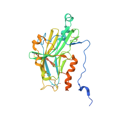
1L2G

Structure of a C-terminally truncated form of glycoprotein D from HSV-1


Experimental Data Snapshot

  • Method: X-RAY DIFFRACTION
  • Resolution: 2.85 Å
  • R-Value Free: 0.297 
  • R-Value Work: 0.278 
  • R-Value Observed: 0.283 

Starting Model: experimental
View more details

wwPDB Validation   3D Report Full Report


Ligand Structure Quality Assessment 


This is version 1.5 of the entry. See complete history


Literature

Herpes simplex virus glycoprotein D bound to the human receptor HveA.

Carfi, A.Willis, S.H.Whitbeck, J.C.Krummenacher, C.Cohen, G.H.Eisenberg, R.J.Wiley, D.C.

(2001) Mol Cell 8: 169-179

  • DOI: https://doi.org/10.1016/s1097-2765(01)00298-2
  • Primary Citation of Related Structures:  
    1JMA, 1L2G

  • PubMed Abstract: 

    Herpes simplex virus (HSV) infection requires binding of the viral envelope glycoprotein D (gD) to cell surface receptors. We report the X-ray structures of a soluble, truncated ectodomain of gD both alone and in complex with the ectodomain of its cellular receptor HveA. Two bound anions suggest possible binding sites for another gD receptor, a 3-O-sulfonated heparan sulfate. Unexpectedly, the structures reveal a V-like immunoglobulin (Ig) fold at the core of gD that is closely related to cellular adhesion molecules and flanked by large N- and C-terminal extensions. The receptor binding segment of gD, an N-terminal hairpin, appears conformationally flexible, suggesting that a conformational change accompanying binding might be part of the viral entry mechanism.


  • Organizational Affiliation

    Department of Medicine, Children's Hospital, Howard Hughes Medical Institute, 320 Longwood Avenue, Boston, MA 02115, USA.


Macromolecules
Find similar proteins by:  (by identity cutoff)  |  3D Structure
Entity ID: 1
MoleculeChains Sequence LengthOrganismDetailsImage
Glycoprotein DA,
B,
C [auth D],
D [auth C]
287Human alphaherpesvirus 1Mutation(s): 0 
UniProt
Find proteins for P57083 (Human herpesvirus 1 (strain Patton))
Explore P57083 
Go to UniProtKB:  P57083
Entity Groups  
Sequence Clusters30% Identity50% Identity70% Identity90% Identity95% Identity100% Identity
UniProt GroupP57083
Glycosylation
Glycosylation Sites: 1
Sequence Annotations
Expand
  • Reference Sequence
Experimental Data & Validation

Experimental Data

  • Method: X-RAY DIFFRACTION
  • Resolution: 2.85 Å
  • R-Value Free: 0.297 
  • R-Value Work: 0.278 
  • R-Value Observed: 0.283 
  • Space Group: P 4
Unit Cell:
Length ( Å )Angle ( ˚ )
a = 131.416α = 90
b = 131.416β = 90
c = 83.265γ = 90
Software Package:
Software NamePurpose
DENZOdata reduction
TRUNCATEdata reduction
AMoREphasing
CNSrefinement
CCP4data scaling

Structure Validation

View Full Validation Report


Ligand Structure Quality Assessment 


Entry History 

Deposition Data

Revision History  (Full details and data files)

  • Version 1.0: 2003-12-16
    Type: Initial release
  • Version 1.1: 2008-04-28
    Changes: Version format compliance
  • Version 1.2: 2011-07-13
    Changes: Non-polymer description, Version format compliance
  • Version 1.3: 2018-05-30
    Changes: Data collection, Derived calculations
  • Version 1.4: 2020-07-29
    Type: Remediation
    Reason: Carbohydrate remediation
    Changes: Data collection, Database references, Derived calculations, Structure summary
  • Version 1.5: 2023-08-16
    Changes: Data collection, Database references, Refinement description, Structure summary