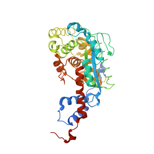
Capture of a labile substrate by expulsion of water molecules from the active site of nicotinate mononucleotide:5,6-dimethylbenzimidazole phosphoribosyltransferase (CobT) from Salmonella enterica.
Cheong, C.G., Escalante-Semerena, J.C., Rayment, I.(2002) J Biological Chem 277: 41120-41127
- PubMed: 12101181
- DOI: https://doi.org/10.1074/jbc.M203535200
- Primary Citation of Related Structures:
1L4B, 1L4E, 1L4F, 1L4G, 1L4H, 1L4K, 1L4L, 1L4M, 1L5O - PubMed Abstract:
Nicotinate mononucleotide (NaMN):5,6-dimethylbenzimidazole (DMB) phosphoribosyltransferase (CobT) from Salmonella enterica plays a central role in the synthesis of alpha-ribazole-5'-phosphate, an intermediate for the lower ligand of cobalamin. In earlier studies it proved difficult to obtain the structure of CobT bound to NaMN because it is hydrolyzed in the crystal lattice in the absence of the second substrate DMB. In an effort to map the reaction pathway of this enzyme, NaMN was captured in the active site with the substrate analogs 4,5-dimethyl-1,2-phenylenediamine, 4-methylcatechol, indole, 3,4-dimethylaniline, 2,5-dimethylaniline, 3,4-dimethylphenol, and 2-amino-p-cresol. Structures of these complexes reveal that they exclude water molecules responsible for the hydrolysis from the active site. These structures, together with the early complexes with alpha-ribazole-5'-phosphate and DMB, provide a complete description of the reaction pathway. They demonstrate that the nicotinate moiety and phosphate do not appear to move significantly between reactants and products but that the aromatic base and ribose moiety each move approximately 1.2 A toward each other in the transformation. This study also reveals that, like many other nucleotide binding proteins, coordination of DMB is accompanied by a disorder-order transition in a surface loop. The structure of apo-CobT is also reported.
- Department of Biochemistry, University of Wisconsin, 433 Babcock Drive, Madison, WI 53706, USA.
Organizational Affiliation:


