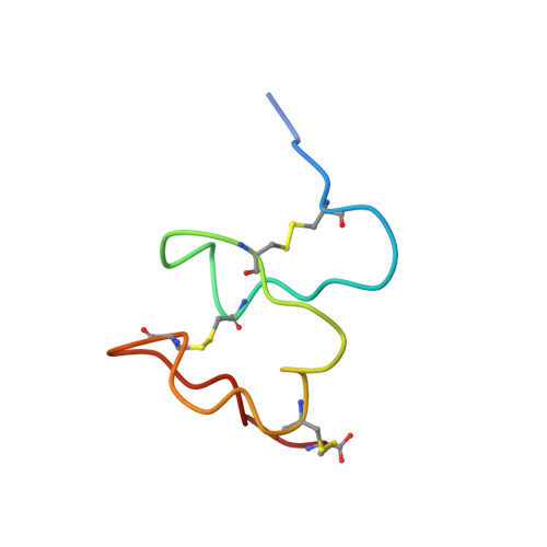
Three-dimensional structure of the second cysteine-rich repeat from the human low-density lipoprotein receptor.
Daly, N.L., Djordjevic, J.T., Kroon, P.A., Smith, R.(1995) Biochemistry 34: 14474-14481
- PubMed: 7578052 Search on PubMed
- DOI: https://doi.org/10.1021/bi00044a025
- Primary Citation Related Structures:
1LDR - PubMed Abstract:
The ligand-binding domain of the low-density lipoprotein receptor comprises seven cysteine-rich repeats, which have been highly conserved through evolution. This domain mediates interactions of the receptor with two lipoprotein apoproteins, apo E and apo B-100, putatively through a calcium-dependent association of the ligands with a cluster of acidic residues on the receptor. The second repeat (rLB2) of the receptor binding domain has been expressed as a thrombin-cleavable GST fusion protein, cleaved, and purified. On oxidation the protein refolded to give a single peak on reverse-phase HPLC. The aqueous solution structure of rLB2 has been determined using two-dimensional 1H NMR spectroscopy. In contrast to the amino-terminal repeat, rLB1, rLB2 has a very flexible structure in water. However, the conformation of rLB2 is markedly more ordered in the presence of a 4-fold molar excess of calcium chloride; the proton resonance dispersion and the number of NOESY cross-peaks are greatly enhanced. The three-dimensional structure of rLB2, obtained from the NMR data by molecular geometry and restrained molecular dynamics methods, parallels that of rLB1, with an amino-terminal hairpin structure followed by a succession of turns. However, there are clear differences in the backbone topology and structural flexibility. As for rLB1, the acidic residues are clustered on one face of the module. The side chain of Asp 37, which is part of a completely conserved SDE sequence thought to be involved in ligand binding, is buried, as is its counterpart (Asp 36) in rLB1. These results provide the first experimental support for the hypothesis that each of the repeats in the ligand-binding domain has a similar global fold but also highlight significant differences in structure and internal dynamics.
- Biochemistry Department, University of Queensland, Australia.
Organizational Affiliation: