Salt bridge relay triggers defective LDL receptor binding by a mutant apolipoprotein.
Wilson, C., Mau, T., Weisgraber, K.H., Wardell, M.R., Mahley, R.W., Agard, D.A.(1994) Structure 2: 713-718
- PubMed: 7994571 Search on PubMed
- DOI: https://doi.org/10.1016/s0969-2126(00)00072-1
- Primary Citation Related Structures:
1LE2 - PubMed Abstract:
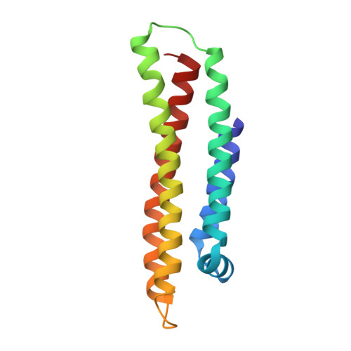
Apolipoprotein-E (apo-E), a 34kDa blood plasma protein, plays a key role in directing cholesterol transport via its interaction with the low density lipoprotein (LDL) receptor. The amino-terminal domain of apo-E forms an unusually elongated four-helix bundle arranged such that key basic residues involved in LDL receptor binding form a cluster at the end of one of the helices. A common apo-E variant, apo-E2, corresponding to the single-site substitution Arg158-->Cys, displays minimal LDL receptor binding and is associated with significant changes in plasma cholesterol levels and increased risk of coronary heart disease. Surprisingly, the site of mutation in this variant is physically well removed (> 12A) from the cluster of LDL receptor binding residues. We now report the refined crystal structure of the amino-terminal domain of apo-E2, at a nominal resolution of 3.0A. This structure reveals significant conformational changes relative to the wild-type protein that may account for reduced LDL receptor binding. Removal of the Arg158 side chain directly disrupts a pair of salt bridges, causing a compensatory reorganization of salt bridge partners that dramatically alters the charge surface presented by apo-E to its receptor. It is proposed that the observed reorganization of surface salt bridges is responsible for the decreased receptor binding by apo-E2. This reorganization, essentially functioning as a mutationally induced electrostatic switch to turn off receptor binding, represents a novel mechanism for the propagation of conformational changes over significant distances.
- Howard Hughes Medical Institute, Department of Biochemistry and Biophysics, University of California, San Francisco 94143-0448.
Organizational Affiliation: