1LJ2 | pdb_00001lj2

Recognition of eIF4G by Rotavirus NSP3 reveals a basis for mRNA circularization


Experimental Data Snapshot

  • Method: X-RAY DIFFRACTION
  • Resolution: 2.38 Å
  • R-Value Free: 
    0.272 (Depositor) 
  • R-Value Work: 
    0.221 (Depositor) 

wwPDB Validation 3D Report Full Report

Validation slider image for 1LJ2

This is version 1.4 of the entry. See complete history

Literature

Recognition of eIF4G by rotavirus NSP3 reveals a basis for mRNA circularization.

Groft, C.M.Burley, S.K.

(2002) Mol Cell 9: 1273-1283

  • DOI: https://doi.org/10.1016/s1097-2765(02)00555-5
  • Primary Citation Related Structures: 
    1LJ2

  • PubMed Abstract: 

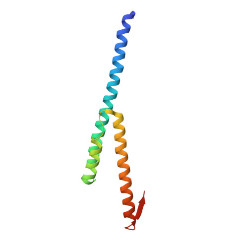
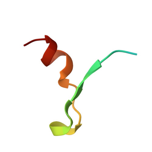
    Rotaviruses, segmented double-stranded RNA viruses, co-opt the eukaryotic translation machinery with the aid of nonstructural protein 3 (NSP3), a rotaviral functional homolog of the cellular poly(A) binding protein (PABP). NSP3 binds to viral mRNA 3' consensus sequences and circularizes mRNA via interactions with eIF4G. Here, we present the X-ray structure of the C-terminal domain of NSP3 (NSP3-C) recognizing a fragment of eIF4GI. Homodimerization of NSP3-C yields a symmetric, elongated, largely alpha-helical structure with two hydrophobic eIF4G binding pockets at the dimer interface. Site-directed mutagenesis and isothermal titration calorimetry documented that NSP3 and PABP use analogous eIF4G recognition strategies, despite marked differences in tertiary structure.


  • Organizational Affiliation
    • Laboratory of Molecular Biophysics, New York, NY 10021, USA.

Macromolecule Content 

  • Total Structure Weight: 32.68 kDa 
  • Atom Count: 2,327 
  • Modeled Residue Count: 265 
  • Deposited Residue Count: 276 
  • Unique protein chains: 2

Macromolecules

Find similar proteins by:|  3D Structure
Entity ID: 1
MoleculeChains  Sequence LengthOrganismDetailsImage
NONSTRUCTURAL RNA-BINDING PROTEIN 34
A, B
110Simian rotavirus A/SA11Mutation(s): 0 
UniProt
Find proteins for P03536 (Rotavirus A (strain RVA/SA11-Both/G3P5B[2]))
Explore P03536 
Go to UniProtKB:  P03536
Entity Groups
Sequence Clusters30% Identity50% Identity70% Identity90% Identity95% Identity100% Identity
UniProt GroupP03536
Sequence Annotations
Expand
Reference Sequence
Find similar proteins by:|  3D Structure
Entity ID: 2
MoleculeChains  Sequence LengthOrganismDetailsImage
eukaryotic protein synthesis initiation factor
C, D
28N/AMutation(s): 0 
UniProt & NIH Common Fund Data Resources
Find proteins for Q04637 (Homo sapiens)
Explore Q04637 
Go to UniProtKB:  Q04637
PHAROS:  Q04637
GTEx:  ENSG00000114867 
Entity Groups
Sequence Clusters30% Identity50% Identity70% Identity90% Identity95% Identity100% Identity
UniProt GroupQ04637
Sequence Annotations
Expand
Reference Sequence

Experimental Data & Validation

Experimental Data

  • Method: X-RAY DIFFRACTION
  • Resolution: 2.38 Å
  • R-Value Free:  0.272 (Depositor) 
  • R-Value Work:  0.221 (Depositor) 
Space Group: P 21 21 21
Unit Cell:
Length ( Å )Angle ( ˚ )
a = 59.69α = 90
b = 74.04β = 90
c = 77.46γ = 90
Software Package:
Software NamePurpose
DENZOdata reduction
TRUNCATEdata reduction
MLPHAREphasing
CNSrefinement
CCP4data scaling

Structure Validation

View Full Validation Report


Entry History 

Deposition Data

Revision History  (Full details and data files)

  • Version 1.0: 2002-07-05
    Type: Initial release
  • Version 1.1: 2008-04-28
    Changes: Version format compliance
  • Version 1.2: 2011-07-13
    Changes: Version format compliance
  • Version 1.3: 2021-02-03
    Changes: Database references, Derived calculations, Structure summary
  • Version 1.4: 2024-02-14
    Changes: Data collection, Database references