1LZV | pdb_00001lzv

Site-Specific Mutant (Tyr7 replaced with His) of Human Carbonic Anhydrase II


Experimental Data Snapshot

  • Method: X-RAY DIFFRACTION
  • Resolution: 2.30 Å
  • R-Value Free: 
    0.251 (Depositor), 0.247 (DCC) 
  • R-Value Work: 
    0.186 (Depositor), 0.184 (DCC) 

Starting Model: experimental
View more details

wwPDB Validation 3D Report Full Report

Validation slider image for 1LZV

This is version 1.4 of the entry. See complete history

Literature

Kinetic analysis of multiple proton shuttles in the active site of human carbonic anhydrase.

Tu, C.Qian, M.An, H.Wadhwa, N.R.Duda, D.Yoshioka, C.Pathak, Y.McKenna, R.Laipis, P.J.Silverman, D.N.

(2002) J Biological Chem 277: 38870-38876

  • DOI: https://doi.org/10.1074/jbc.M205791200
  • Primary Citation Related Structures: 
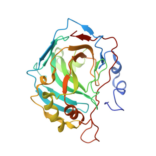
    1LZV

  • PubMed Abstract: 

    We have prepared a site-specific mutant of human carbonic anhydrase (HCA) II with histidine residues at positions 7 and 64 in the active site cavity. Using a different isozyme, we have placed histidine residues in HCA III at positions 64 and 67 and in another mutant at positions 64 and 7. Each of these histidine residues can act as a proton transfer group in catalysis when it is the only nonliganding histidine in the active site cavity, except His(7) in HCA III. Using an (18)O exchange method to measure rate constants for intramolecular proton transfer, we have found that inserting two histidine residues into the active site cavity of either isozyme II or III of carbonic anhydrase results in rates of proton transfer to the zinc-bound hydroxide that are antagonistic or suppressive with respect to the corresponding single mutants. The crystal structure of Y7H HCA II, which contains both His(7) and His(64) within the active site cavity, shows the conformation of the side chain of His(64) moved from its position in the wild type and hydrogen-bonded through an intervening water molecule with the side chain of His(7). This suggests a cause of decreased proton transfer in catalysis.


  • Organizational Affiliation
    • Department of Pharmacology and Therapeutics, University of Florida College of Medicine, Gainesville, Florida 32610-0267, USA.

Macromolecule Content 

  • Total Structure Weight: 29.33 kDa 
  • Atom Count: 2,093 
  • Modeled Residue Count: 256 
  • Deposited Residue Count: 260 
  • Unique protein chains: 1

Macromolecules

Find similar proteins by:|  3D Structure
Entity ID: 1
MoleculeChains  Sequence LengthOrganismDetailsImage
Carbonic Anhydrase II260Homo sapiensMutation(s): 1 
EC: 4.2.1.1 (PDB Primary Data), 4.2.1.69 (UniProt)
UniProt & NIH Common Fund Data Resources
Find proteins for P00918 (Homo sapiens)
Explore P00918 
Go to UniProtKB:  P00918
PHAROS:  P00918
GTEx:  ENSG00000104267 
Entity Groups
Sequence Clusters30% Identity50% Identity70% Identity90% Identity95% Identity100% Identity
UniProt GroupP00918
Sequence Annotations
Expand
Reference Sequence

Small Molecules

Ligands 1 Unique
IDChains Name / Formula / InChI Key2D Diagram3D Interactions
ZN

Query on ZN


Download:Ideal Coordinates CCD File
B [auth A]ZINC ION
Zn
PTFCDOFLOPIGGS-UHFFFAOYSA-N

Experimental Data & Validation

Experimental Data

  • Method: X-RAY DIFFRACTION
  • Resolution: 2.30 Å
  • R-Value Free:  0.251 (Depositor), 0.247 (DCC) 
  • R-Value Work:  0.186 (Depositor), 0.184 (DCC) 
Space Group: P 1 21 1
Unit Cell:
Length ( Å )Angle ( ˚ )
a = 42.871α = 90
b = 41.585β = 104.75
c = 72.951γ = 90
Software Package:
Software NamePurpose
DENZOdata reduction
SCALEPACKdata scaling
CNSrefinement
CNSphasing

Structure Validation

View Full Validation Report


Entry History 

Deposition Data

Revision History  (Full details and data files)

  • Version 1.0: 2002-10-23
    Type: Initial release
  • Version 1.1: 2008-04-28
    Changes: Version format compliance
  • Version 1.2: 2011-07-13
    Changes: Version format compliance
  • Version 1.3: 2021-10-27
    Changes: Database references, Derived calculations
  • Version 1.4: 2024-02-14
    Changes: Data collection, Refinement description