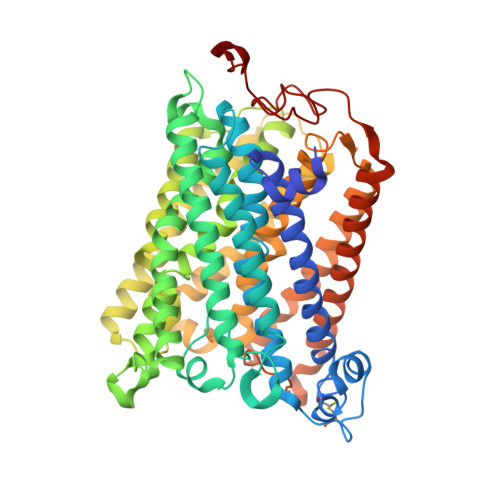
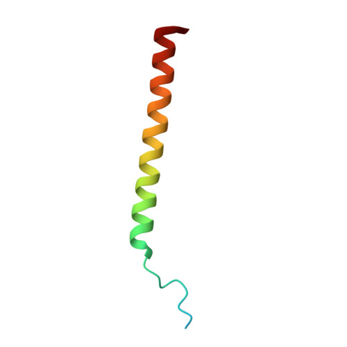
1M57

Structure of cytochrome c oxidase from Rhodobacter sphaeroides (EQ(I-286) mutant))


Experimental Data Snapshot

  • Method: X-RAY DIFFRACTION
  • Resolution: 3.00 Å
  • R-Value Free: 0.329 
  • R-Value Work: 0.293 
  • R-Value Observed: 0.293 

wwPDB Validation   3D Report Full Report


This is version 2.2 of the entry. See complete history


Literature

The X-ray crystal structures of wild-type and EQ(I-286) mutant cytochrome c oxidases from Rhodobacter sphaeroides.

Svensson-Ek, M.Abramson, J.Larsson, G.Tornroth, S.Brzezinski, P.Iwata, S.

(2002) J Mol Biol 321: 329-339

  • DOI: https://doi.org/10.1016/s0022-2836(02)00619-8
  • Primary Citation of Related Structures:  
    1M56, 1M57

  • PubMed Abstract: 

    The structure of cytochrome c oxidase from Rhodobacter sphaeroides has been solved at 2.3/2.8A (anisotropic resolution). This high-resolution structure revealed atomic details of a bacterial terminal oxidase including water molecule positions and a potential oxygen pathway, which has not been reported in other oxidase structures. A comparative study of the wild-type and the EQ(I-286) mutant enzyme revealed structural rearrangements around E(I-286) that could be crucial for proton transfer in this enzyme. In the structure of the mutant enzyme, EQ(I-286), which cannot transfer protons during oxygen reduction, the side-chain of Q(I-286) does not have the hydrogen bond to the carbonyl oxygen of M(I-107) that is seen in the wild-type structure. Furthermore, the Q(I-286) mutant has a different arrangement of water molecules and residues in the vicinity of the Q side-chain. These differences between the structures could reflect conformational changes that take place upon deprotonation of E(I-286) during turnover of the wild-type enzyme, which could be part of the proton-pumping machinery of the enzyme.


  • Organizational Affiliation

    Department of Biochemistry, Biomedical Center, Uppsala University, Box 576, Uppsala, Sweden. margareta.e.ek@astrazeneca.com


Macromolecules
Find similar proteins by:  (by identity cutoff)  |  3D Structure
Entity ID: 1
MoleculeChains Sequence LengthOrganismDetailsImage
CYTOCHROME C OXIDASEA,
E [auth G]
566Cereibacter sphaeroidesMutation(s): 1 
EC: 1.9.3.1 (PDB Primary Data), 7.1.1.9 (UniProt)
Membrane Entity: Yes 
UniProt
Find proteins for P33517 (Cereibacter sphaeroides)
Explore P33517 
Go to UniProtKB:  P33517
Entity Groups  
Sequence Clusters30% Identity50% Identity70% Identity90% Identity95% Identity100% Identity
UniProt GroupP33517
Sequence Annotations
Expand
  • Reference Sequence
Find similar proteins by:  (by identity cutoff)  |  3D Structure
Entity ID: 2
MoleculeChains Sequence LengthOrganismDetailsImage
CYTOCHROME C OXIDASEB,
F [auth H]
264Cereibacter sphaeroidesMutation(s): 0 
EC: 1.9.3.1 (PDB Primary Data), 7.1.1.9 (UniProt)
Membrane Entity: Yes 
UniProt
Find proteins for Q03736 (Cereibacter sphaeroides)
Explore Q03736 
Go to UniProtKB:  Q03736
Entity Groups  
Sequence Clusters30% Identity50% Identity70% Identity90% Identity95% Identity100% Identity
UniProt GroupQ03736
Sequence Annotations
Expand
  • Reference Sequence
Find similar proteins by:  (by identity cutoff)  |  3D Structure
Entity ID: 3
MoleculeChains Sequence LengthOrganismDetailsImage
CYTOCHROME C OXIDASEC,
G [auth I]
266Cereibacter sphaeroidesMutation(s): 0 
EC: 1.9.3.1 (PDB Primary Data), 7.1.1.9 (UniProt)
Membrane Entity: Yes 
UniProt
Find proteins for P84153 (Cereibacter sphaeroides)
Explore P84153 
Go to UniProtKB:  P84153
Entity Groups  
Sequence Clusters30% Identity50% Identity70% Identity90% Identity95% Identity100% Identity
UniProt GroupP84153
Sequence Annotations
Expand
  • Reference Sequence
Find similar proteins by:  (by identity cutoff)  |  3D Structure
Entity ID: 4
MoleculeChains Sequence LengthOrganismDetailsImage
CYTOCHROME C OXIDASED,
H [auth J]
51Cereibacter sphaeroidesMutation(s): 0 
EC: 1.9.3.1
Membrane Entity: Yes 
UniProt
Find proteins for Q8KRK5 (Cereibacter sphaeroides)
Explore Q8KRK5 
Go to UniProtKB:  Q8KRK5
Entity Groups  
Sequence Clusters30% Identity50% Identity70% Identity90% Identity95% Identity100% Identity
UniProt GroupQ8KRK5
Sequence Annotations
Expand
  • Reference Sequence
Small Molecules
Ligands 5 Unique
IDChains Name / Formula / InChI Key2D Diagram3D Interactions
HEA
Query on HEA

Download Ideal Coordinates CCD File 
L [auth A],
M [auth A],
Y [auth G],
Z [auth G]
HEME-A
C49 H56 Fe N4 O6
ZGGYGTCPXNDTRV-PRYGPKJJSA-L
3PE
Query on 3PE

Download Ideal Coordinates CCD File 
AA [auth G]
BA [auth G]
EA [auth I]
FA [auth I]
GA [auth I]
AA [auth G],
BA [auth G],
EA [auth I],
FA [auth I],
GA [auth I],
HA [auth J],
N [auth A],
O [auth A],
R [auth C],
S [auth C],
T [auth C],
U [auth D]
1,2-Distearoyl-sn-glycerophosphoethanolamine
C41 H82 N O8 P
LVNGJLRDBYCPGB-LDLOPFEMSA-N
CU
Query on CU

Download Ideal Coordinates CCD File 
CA [auth H]
DA [auth H]
I [auth A]
P [auth B]
Q [auth B]
CA [auth H],
DA [auth H],
I [auth A],
P [auth B],
Q [auth B],
V [auth G]
COPPER (II) ION
Cu
JPVYNHNXODAKFH-UHFFFAOYSA-N
CA
Query on CA

Download Ideal Coordinates CCD File 
K [auth A],
X [auth G]
CALCIUM ION
Ca
BHPQYMZQTOCNFJ-UHFFFAOYSA-N
MG
Query on MG

Download Ideal Coordinates CCD File 
J [auth A],
W [auth G]
MAGNESIUM ION
Mg
JLVVSXFLKOJNIY-UHFFFAOYSA-N
Experimental Data & Validation

Experimental Data

  • Method: X-RAY DIFFRACTION
  • Resolution: 3.00 Å
  • R-Value Free: 0.329 
  • R-Value Work: 0.293 
  • R-Value Observed: 0.293 
  • Space Group: H 3
Unit Cell:
Length ( Å )Angle ( ˚ )
a = 340.72α = 90
b = 340.72β = 90
c = 89.76γ = 120
Software Package:
Software NamePurpose
DENZOdata reduction
SCALEPACKdata scaling
AMoREphasing
REFMACrefinement

Structure Validation

View Full Validation Report


Entry History 

Deposition Data

Revision History  (Full details and data files)

  • Version 1.0: 2002-08-28
    Type: Initial release
  • Version 1.1: 2008-04-28
    Changes: Version format compliance
  • Version 1.2: 2011-07-13
    Changes: Version format compliance
  • Version 2.0: 2021-06-30
    Changes: Atomic model, Data collection, Derived calculations, Non-polymer description, Structure summary
  • Version 2.1: 2021-11-10
    Changes: Database references
  • Version 2.2: 2024-10-30
    Changes: Data collection, Structure summary