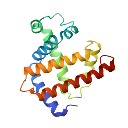
X-ray structure and refinement of carbon-monoxy (Fe II)-myoglobin at 1.5 A resolution.
Kuriyan, J., Wilz, S., Karplus, M., Petsko, G.A.(1986) J Mol Biology 192: 133-154
- PubMed: 3820301
- DOI: https://doi.org/10.1016/0022-2836(86)90470-5
- Primary Citation of Related Structures:
1MBC - PubMed Abstract:
The structure of carbon-monoxy (Fe II) myoglobin at 260 K has been solved at a resolution of 1.5 A by X-ray diffraction and a model refined against the X-ray data by restrained least-squares. The CO ligand is disordered and distorted from the linear conformation seen in model compounds. At least two conformations, with Fe--C--O angles of 140 degrees and 120 degrees, are required to model the system. The heme pocket is significantly larger than in deoxy-myoglobin because the distal residues have relaxed around the ligand; the largest displacement occurs for the distal histidine side-chain, which moves more than 1.4 A on ligand binding. The side-chain of Arg45 (CD3) is disordered and apparently exists in two equally populated conformations. One of these does not block the motion of the distal histidine out of the binding pocket, suggesting a mechanism for ligand entry. The heme group is planar (root-mean-square deviation from planarity is 0.08 A) with no doming of the pyrrole groups. The Fe--N epsilon 2 (His93) bond length is 2.2 A and the Fe--C bond length in the CO complex is 1.9 A. The iron is the least-squares plane of the heme, and this leads to the proximal histidine moving by 0.4 A relative to its position in deoxy-myoglobin. This shift correlates with a global structural change, with the proximal part of the molecule translated towards the heme plane.


