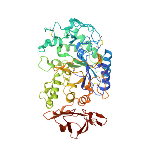
1MFV

Probing the role of a mobile loop in human slaivary amylase: Structural studies on the loop-deleted enzyme


Experimental Data Snapshot

  • Method: X-RAY DIFFRACTION
  • Resolution: 2.00 Å
  • R-Value Free: 0.195 
  • R-Value Work: 0.167 
  • R-Value Observed: 0.169 

Starting Model: experimental
View more details

wwPDB Validation   3D Report Full Report


Ligand Structure Quality Assessment 


This is version 3.1 of the entry. See complete history


Literature

Probing the role of a mobile loop in substrate binding and enzyme activity of human salivary amylase.

Ramasubbu, N.Ragunath, C.Mishra, P.J.

(2003) J Mol Biol 325: 1061-1076

  • DOI: https://doi.org/10.1016/s0022-2836(02)01326-8
  • Primary Citation of Related Structures:  
    1JXK, 1MFU, 1MFV

  • PubMed Abstract: 

    Mammalian amylases harbor a flexible, glycine-rich loop 304GHGAGGA(310), which becomes ordered upon oligosaccharide binding and moves in toward the substrate. In order to probe the role of this loop in catalysis, a deletion mutant lacking residues 306-310 (Delta306) was generated. Kinetic studies showed that Delta306 exhibited: (1) a reduction (>200-fold) in the specific activity using starch as a substrate; (2) a reduction in k(cat) for maltopentaose and maltoheptaose as substrates; and (3) a twofold increase in K(m) (maltopentaose as substrate) compared to the wild-type (rHSAmy). More cleavage sites were observed for the mutant than for rHSAmy, suggesting that the mutant exhibits additional productive binding modes. Further insight into its role is obtained from the crystal structures of the two enzymes soaked with acarbose, a transition-state analog. Both enzymes modify acarbose upon binding through hydrolysis, condensation or transglycosylation reactions. Electron density corresponding to six and seven fully occupied subsites in the active site of rHSAmy and Delta306, respectively, were observed. Comparison of the crystal structures showed that: (1) the hydrophobic cover provided by the mobile loop for the subsites at the reducing end of the rHSAmy complex is notably absent in the mutant; (2) minimal changes in the protein-ligand interactions around subsites S1 and S1', where the cleavage would occur; (3) a well-positioned water molecule in the mutant provides a hydrogen bond interaction similar to that provided by the His305 in rHSAmy complex; (4) the active site-bound oligosaccharides exhibit minimal conformational differences between the two enzymes. Collectively, while the kinetic data suggest that the mobile loop may be involved in assisting the catalysis during the transition state, crystallographic data suggest that the loop may play a role in the release of the product(s) from the active site.


  • Organizational Affiliation

    Department of Oral Biology, University of Medicine and Dentistry of New Jersey, Newark, NJ 07103, USA. n.ramasubbu@umdnj.edu


Macromolecules
Find similar proteins by:  (by identity cutoff)  |  3D Structure
Entity ID: 1
MoleculeChains Sequence LengthOrganismDetailsImage
alpha-amylase, salivary496Homo sapiensMutation(s): 0 
EC: 3.2.1.1
UniProt & NIH Common Fund Data Resources
Find proteins for P0DUB6 (Homo sapiens)
Explore P0DUB6 
Go to UniProtKB:  P0DUB6
GTEx:  ENSG00000237763 
Entity Groups  
Sequence Clusters30% Identity50% Identity70% Identity90% Identity95% Identity100% Identity
UniProt GroupP0DUB6
Sequence Annotations
Expand
  • Reference Sequence
Oligosaccharides

Help

Entity ID: 2
MoleculeChains Length2D Diagram Glycosylation3D Interactions
4-amino-4,6-dideoxy-alpha-D-glucopyranose-(1-4)-alpha-D-glucopyranose
B, C
2N/A
Glycosylation Resources
GlyTouCan:  G90989WB
GlyCosmos:  G90989WB
Entity ID: 3
MoleculeChains Length2D Diagram Glycosylation3D Interactions
4-amino-4,6-dideoxy-alpha-D-glucopyranose-(1-4)-alpha-D-glucopyranose-(1-4)-beta-D-glucopyranose
D
3N/A
Glycosylation Resources
GlyTouCan:  G21221NT
GlyCosmos:  G21221NT
Experimental Data & Validation

Experimental Data

  • Method: X-RAY DIFFRACTION
  • Resolution: 2.00 Å
  • R-Value Free: 0.195 
  • R-Value Work: 0.167 
  • R-Value Observed: 0.169 
  • Space Group: P 21 21 21
Unit Cell:
Length ( Å )Angle ( ˚ )
a = 51.681α = 90
b = 73.197β = 90
c = 134.448γ = 90
Software Package:
Software NamePurpose
DENZOdata reduction
SCALEPACKdata scaling
AMoREphasing
REFMACrefinement

Structure Validation

View Full Validation Report


Ligand Structure Quality Assessment 


Entry History 

Deposition Data

Revision History  (Full details and data files)

  • Version 1.0: 2002-11-20
    Type: Initial release
  • Version 1.1: 2008-04-28
    Changes: Version format compliance
  • Version 1.2: 2011-07-13
    Changes: Version format compliance
  • Version 2.0: 2019-12-25
    Changes: Data collection, Database references, Derived calculations, Polymer sequence
  • Version 3.0: 2020-07-29
    Type: Remediation
    Reason: Carbohydrate remediation
    Changes: Atomic model, Data collection, Derived calculations, Structure summary
  • Version 3.1: 2024-10-30
    Changes: Data collection, Database references, Derived calculations, Refinement description, Structure summary