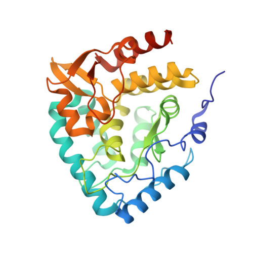
1MLW

Crystal structure of human tryptophan hydroxylase with bound 7,8-dihydro-L-biopterin cofactor and Fe(III)


Experimental Data Snapshot

  • Method: X-RAY DIFFRACTION
  • Resolution: 1.71 Å
  • R-Value Free: 0.230 
  • R-Value Work: 0.205 

Starting Model: experimental
View more details

wwPDB Validation   3D Report Full Report


Ligand Structure Quality Assessment 


This is version 1.3 of the entry. See complete history


Literature

Three-dimensional structure of human tryptophan hydroxylase and its implications for the biosynthesis of the neurotransmitters serotonin and melatonin

Wang, L.Erlandsen, H.Haavik, J.Knappskog, P.M.Stevens, R.C.

(2002) Biochemistry 41: 12569-12574

  • DOI: https://doi.org/10.1021/bi026561f
  • Primary Citation of Related Structures:  
    1MLW

  • PubMed Abstract: 

    Tryptophan hydroxylase oxidizes L-tryptophan to 5-hydroxy-L-tryptophan in the rate-determining step of serotonin biosynthesis. We have determined the X-ray crystal structure (1.7 A) of a truncated functional form of human tryptophan hydroxylase with the bound cofactor analogue 7,8-dihydro-L-biopterin, providing the first atomic-resolution information for the catalytic domain of this important enzyme. Comparison of the three-dimensional structures of all three members of the aromatic amino acid hydroxylase family--tyrosine hydroxylase, phenylalanine hydroxylase, and tryptophan hydroxylase--reveals important differences at the active sites.


  • Organizational Affiliation

    Department of Molecular Biology, The Scripps Research Institute, 10550 North Torrey Pines Road, La Jolla, California 92037, USA.


Macromolecules
Find similar proteins by:  (by identity cutoff)  |  3D Structure
Entity ID: 1
MoleculeChains Sequence LengthOrganismDetailsImage
Tryptophan 5-monooxygenase301Homo sapiensMutation(s): 0 
Gene Names: TPH
EC: 1.14.16.4
UniProt & NIH Common Fund Data Resources
Find proteins for P17752 (Homo sapiens)
Explore P17752 
Go to UniProtKB:  P17752
PHAROS:  P17752
GTEx:  ENSG00000129167 
Entity Groups  
Sequence Clusters30% Identity50% Identity70% Identity90% Identity95% Identity100% Identity
UniProt GroupP17752
Sequence Annotations
Expand
  • Reference Sequence
Experimental Data & Validation

Experimental Data

  • Method: X-RAY DIFFRACTION
  • Resolution: 1.71 Å
  • R-Value Free: 0.230 
  • R-Value Work: 0.205 
  • Space Group: P 21 21 21
Unit Cell:
Length ( Å )Angle ( ˚ )
a = 47.775α = 90
b = 57.334β = 90
c = 109.41γ = 90
Software Package:
Software NamePurpose
HKL-2000data collection
SCALEPACKdata scaling
EPMRphasing
CNSrefinement
HKL-2000data reduction

Structure Validation

View Full Validation Report


Ligand Structure Quality Assessment 


Entry History 

Deposition Data

Revision History  (Full details and data files)

  • Version 1.0: 2002-12-18
    Type: Initial release
  • Version 1.1: 2007-10-16
    Changes: Version format compliance
  • Version 1.2: 2011-07-13
    Changes: Version format compliance
  • Version 1.3: 2024-02-14
    Changes: Data collection, Database references, Derived calculations, Refinement description