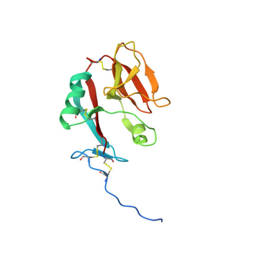
1MPU

Crystal Structure of the free human NKG2D immunoreceptor


Experimental Data Snapshot

  • Method: X-RAY DIFFRACTION
  • Resolution: 2.50 Å
  • R-Value Free: 0.296 
  • R-Value Work: 0.245 

Starting Model: experimental
View more details

wwPDB Validation   3D Report Full Report


This is version 1.6 of the entry. See complete history


Literature

Symmetry Recognizing Asymmetry: Analysis of the Interactions between the C-Type Lectin-like Immunoreceptor NKG2D and MHC Class I-like Ligands

McFarland, B.J.Kortemme, T.Yu, S.F.Baker, D.Strong, R.K.

(2003) Structure 11: 411-422

  • DOI: https://doi.org/10.1016/s0969-2126(03)00047-9
  • Primary Citation of Related Structures:  
    1MPU

  • PubMed Abstract: 

    Engagement of diverse protein ligands (MIC-A/B, ULBP, Rae-1, or H60) by NKG2D immunoreceptors mediates elimination of tumorigenic or virally infected cells by natural killer and T cells. Three previous NKG2D-ligand complex structures show the homodimeric receptor interacting with the monomeric ligands in similar 2:1 complexes, with an equivalent surface on each NKG2D monomer binding intimately to a total of six distinct ligand surfaces. Here, the crystal structure of free human NKG2D and in silico and in vitro alanine-scanning mutagenesis analyses of the complex interfaces indicate that NKG2D recognition degeneracy is not explained by a classical induced-fit mechanism. Rather, the divergent ligands appear to utilize different strategies to interact with structurally conserved elements of the consensus NKG2D binding site.


  • Organizational Affiliation

    The Division of Basic Sciences, Fred Hutchinson Cancer Research Center, 1100 Fairview Avenue North, Seattle, WA 98109, USA.


Macromolecules
Find similar proteins by:  (by identity cutoff)  |  3D Structure
Entity ID: 1
MoleculeChains Sequence LengthOrganismDetailsImage
NKG2-D type II integral membrane protein138Homo sapiensMutation(s): 0 
UniProt & NIH Common Fund Data Resources
Find proteins for P26718 (Homo sapiens)
Explore P26718 
Go to UniProtKB:  P26718
PHAROS:  P26718
GTEx:  ENSG00000213809 
Entity Groups  
Sequence Clusters30% Identity50% Identity70% Identity90% Identity95% Identity100% Identity
UniProt GroupP26718
Sequence Annotations
Expand
  • Reference Sequence
Small Molecules
Ligands 1 Unique
IDChains Name / Formula / InChI Key2D Diagram3D Interactions
PO4
Query on PO4

Download Ideal Coordinates CCD File 
B [auth A]PHOSPHATE ION
O4 P
NBIIXXVUZAFLBC-UHFFFAOYSA-K
Experimental Data & Validation

Experimental Data

  • Method: X-RAY DIFFRACTION
  • Resolution: 2.50 Å
  • R-Value Free: 0.296 
  • R-Value Work: 0.245 
  • Space Group: P 41 21 2
Unit Cell:
Length ( Å )Angle ( ˚ )
a = 87.65α = 90
b = 87.65β = 90
c = 36.13γ = 90
Software Package:
Software NamePurpose
DENZOdata reduction
SCALEPACKdata scaling
EPMRphasing
CNSrefinement

Structure Validation

View Full Validation Report


Entry History 

Deposition Data

Revision History  (Full details and data files)

  • Version 1.0: 2003-04-15
    Type: Initial release
  • Version 1.1: 2008-04-28
    Changes: Version format compliance
  • Version 1.2: 2011-07-13
    Changes: Derived calculations, Version format compliance
  • Version 1.3: 2018-04-04
    Changes: Data collection
  • Version 1.4: 2022-12-21
    Changes: Database references, Derived calculations
  • Version 1.5: 2023-09-20
    Changes: Data collection, Refinement description
  • Version 1.6: 2024-11-13
    Changes: Structure summary