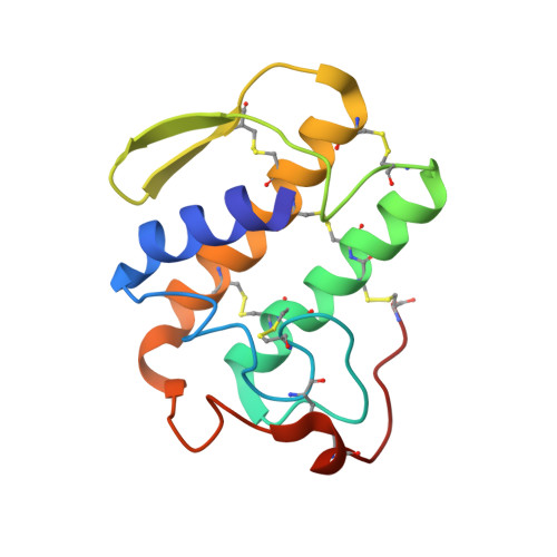
The crystal structure of the H48Q active site mutant of human group IIA secreted phospholipase A2 at 1.5 A resolution provides an insight into the catalytic mechanism
Edwards, S.H., Thompson, D., Baker, S.F., Wood, S.P., Wilton, D.C.(2002) Biochemistry 41: 15468-15476
- PubMed: 12501175
- DOI: https://doi.org/10.1021/bi020485z
- Primary Citation of Related Structures:
1N28, 1N29 - PubMed Abstract:
The human group IIA secreted PLA(2) is a 14 kDa calcium-dependent extracellular enzyme that has been characterized as an acute phase protein with important antimicrobial activity and has been implicated in signal transduction. The selective binding of this enzyme to the phospholipid substrate interface plays a crucial role in its physiological function. To study interfacial binding in the absence of catalysis, one strategy is to produce structurally intact but catalytically inactive mutants. The active site mutants H48Q, H48N, and H48A had been prepared for the secreted PLA(2)s from bovine pancreas and bee venom and retained minimal catalytic activity while the H48Q mutant showed the maximum structural integrity. Preparation of the mutant H48Q of the human group IIA enzyme unexpectedly produced an enzyme that retained significant (2-4%) catalytic activity that was contrary to expectations in view of the accepted catalytic mechanism. In this paper it is established that the high residual activity of the H48Q mutant is genuine, not due to contamination, and can be seen under a variety of assay conditions including assays in the presence of Co(2+) and Ni(2+) in place of Ca(2+). The crystallization of the H48Q mutant, yielding diffraction data to a resolution of 1.5 A, allowed a comparison with the corresponding recombinant wild-type enzyme (N1A) that was also crystallized. This comparison revealed that all of the important features of the catalytic machinery were in place and the two structures were virtually superimposable. In particular, the catalytic calcium ion occupied an identical position in the active site of the two proteins, and the catalytic water molecule (w6) was clearly resolved in the H48Q mutant. We propose that a variation of the calcium-coordinated oxyanion ("two water") mechanism involving hydrogen bonding rather than the anticipated full proton transfer to the histidine will best explain the ability of an active site glutamine to allow significant catalytic activity.
- Division of Biochemistry and Molecular Biology, School of Biological Sciences, University of Southampton, Bassett Crescent East, Southampton SO16 7PX, U.K.
Organizational Affiliation:


