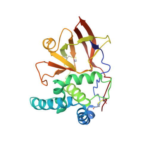
1OG1

CRYSTAL STRUCTURE OF THE EUCARYOTIC MONO-ADP-RIBOSYLTRANSFERASE ART2.2 IN COMPLEX WITH TAD


Experimental Data Snapshot

  • Method: X-RAY DIFFRACTION
  • Resolution: 2.00 Å
  • R-Value Free: 0.244 
  • R-Value Work: 0.202 
  • R-Value Observed: 0.202 

Starting Model: experimental
View more details

wwPDB Validation   3D Report Full Report


Ligand Structure Quality Assessment 


This is version 1.6 of the entry. See complete history


Literature

Substrate Binding and Catalysis of Ecto-Adp-Ribosyltransferase 2.2 From Rat

Ritter, H.Koch-Nolte, F.Marquez, V.E.Schulz, G.E.

(2003) Biochemistry 42: 10155

  • DOI: https://doi.org/10.1021/bi034625w
  • Primary Citation of Related Structures:  
    1OG1, 1OG3, 1OG4

  • PubMed Abstract: 

    The structures of beta-methylenethiazole-4-carboxamide adenine dinucleotide (TAD), NAD(+), and NADH as bound to ecto-ADP-ribosyltransferase 2.2 from rat and to its mutants E189I and E189A, respectively, have been established. The positions and conformations of NAD(+) and its analogues agree in general with those in other ADP-ribosyltransferases. The kinetic constants for NAD(+) hydrolysis were determined by RP-HPLC. The specific activity amounts to 26 units/mg, which is 6000-fold higher than a previously reported rate and 500-fold higher than the hydrolysis rates of other ADP-ribosyltransferases, confirming that hydrolysis is the major function of this enzyme. On the basis of structures and mutant activities, a catalytic mechanism is proposed. The known auto-ADP-ribosylation of the enzyme at the suggested position R184 is supported by one of the crystal structures where the nucleophile position is occupied by an Neta atom of this arginine which in turn is backed up by the base E159.


  • Organizational Affiliation

    Institut für Organische Chemie und Biochemie, Albert-Ludwigs-Universität, Albertstrasse 21, Freiburg im Breisgau 79104, Germany.


Macromolecules
Find similar proteins by:  (by identity cutoff)  |  3D Structure
Entity ID: 1
MoleculeChains Sequence LengthOrganismDetailsImage
T-CELL ECTO-ADP-RIBOSYLTRANSFERASE 2226Rattus norvegicusMutation(s): 0 
EC: 2.4.2.31 (PDB Primary Data), 3.2.2.5 (UniProt)
UniProt
Find proteins for P20974 (Rattus norvegicus)
Explore P20974 
Go to UniProtKB:  P20974
Entity Groups  
Sequence Clusters30% Identity50% Identity70% Identity90% Identity95% Identity100% Identity
UniProt GroupP20974
Sequence Annotations
Expand
  • Reference Sequence
Small Molecules
Ligands 1 Unique
IDChains Name / Formula / InChI Key2D Diagram3D Interactions
TAD
Query on TAD

Download Ideal Coordinates CCD File 
B [auth A]BETA-METHYLENE-THIAZOLE-4-CARBOXYAMIDE-ADENINE DINUCLEOTIDE
C20 H27 N7 O13 P2 S
CRWWKLKZKYLFQV-HVIRUEHBSA-N
Experimental Data & Validation

Experimental Data

  • Method: X-RAY DIFFRACTION
  • Resolution: 2.00 Å
  • R-Value Free: 0.244 
  • R-Value Work: 0.202 
  • R-Value Observed: 0.202 
  • Space Group: P 31 2 1
Unit Cell:
Length ( Å )Angle ( ˚ )
a = 81.419α = 90
b = 81.419β = 90
c = 77.265γ = 120
Software Package:
Software NamePurpose
DENZOdata reduction
SCALEPACKdata scaling
MOLREPphasing
PHENIXphasing
CNSrefinement

Structure Validation

View Full Validation Report


Ligand Structure Quality Assessment 


Entry History 

Deposition Data

Revision History  (Full details and data files)

  • Version 1.0: 2003-08-28
    Type: Initial release
  • Version 1.1: 2011-05-08
    Changes: Version format compliance
  • Version 1.2: 2011-07-13
    Changes: Version format compliance
  • Version 1.3: 2017-07-12
    Changes: Refinement description
  • Version 1.4: 2019-07-24
    Changes: Data collection
  • Version 1.5: 2023-12-13
    Changes: Data collection, Database references, Other, Refinement description
  • Version 1.6: 2024-10-16
    Changes: Structure summary