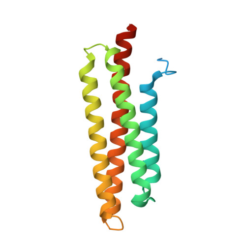
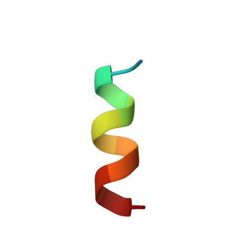
1OW7

Paxillin LD4 motif bound to the Focal Adhesion Targeting (FAT) domain of the Focal Adhesion Kinase


Experimental Data Snapshot

  • Method: X-RAY DIFFRACTION
  • Resolution: 2.60 Å
  • R-Value Free: 0.269 
  • R-Value Work: 0.237 
  • R-Value Observed: 0.239 

Starting Model: experimental
View more details

wwPDB Validation   3D Report Full Report


This is version 1.3 of the entry. See complete history


Literature

Molecular Recognition of Paxillin LD Motifs by the Focal Adhesion Targeting Domain

Hoellerer, M.K.Noble, M.E.M.Labesse, G.Werner, J.M.Arold, S.T.

(2003) Structure 11: 1207-1217

  • DOI: https://doi.org/10.1016/j.str.2003.08.010
  • Primary Citation of Related Structures:  
    1OW6, 1OW7, 1OW8

  • PubMed Abstract: 

    Focal adhesions (FAs) are large submembrane signaling complexes formed at sites of cellular attachment to the extracellular matrix. The interaction of LD motifs with their targets plays an important role in the assembly of FAs. We have determined the molecular basis for the recognition of two paxillin LD motifs by the FA targeting (FAT) domain of FA kinase using a combination of X-ray crystallography, solution NMR, and homology modeling. The four-helix FAT domain displays two LD binding sites on opposite sites of the molecule that bind LD peptides in a helical conformation. Threading studies suggest that the LD-interacting domain of p95PKL shares a common four-helical core with the FAT domain and the tail of vinculin, defining a structural family of LD motif binding modules.


  • Organizational Affiliation

    Biochemistry Department, South Parks Road, Oxford, OX1 3QU, United Kingdom.


Macromolecules
Find similar proteins by:  (by identity cutoff)  |  3D Structure
Entity ID: 1
MoleculeChains Sequence LengthOrganismDetailsImage
Focal adhesion kinase 1
A, B, C
161Homo sapiensMutation(s): 0 
Gene Names: PTK2 OR FAK1 OR FAK
EC: 2.7.1.112 (PDB Primary Data), 2.7.10.2 (UniProt)
UniProt & NIH Common Fund Data Resources
Find proteins for Q05397 (Homo sapiens)
Explore Q05397 
Go to UniProtKB:  Q05397
PHAROS:  Q05397
GTEx:  ENSG00000169398 
Entity Groups  
Sequence Clusters30% Identity50% Identity70% Identity90% Identity95% Identity100% Identity
UniProt GroupQ05397
Sequence Annotations
Expand
  • Reference Sequence

Find similar proteins by:  Sequence   |   3D Structure  

Entity ID: 2
MoleculeChains Sequence LengthOrganismDetailsImage
Paxillin
D, E, F
13N/AMutation(s): 0 
UniProt & NIH Common Fund Data Resources
Find proteins for P49023 (Homo sapiens)
Explore P49023 
Go to UniProtKB:  P49023
PHAROS:  P49023
GTEx:  ENSG00000089159 
Entity Groups  
Sequence Clusters30% Identity50% Identity70% Identity90% Identity95% Identity100% Identity
UniProt GroupP49023
Sequence Annotations
Expand
  • Reference Sequence
Experimental Data & Validation

Experimental Data

  • Method: X-RAY DIFFRACTION
  • Resolution: 2.60 Å
  • R-Value Free: 0.269 
  • R-Value Work: 0.237 
  • R-Value Observed: 0.239 
  • Space Group: C 2 2 21
Unit Cell:
Length ( Å )Angle ( ˚ )
a = 88.994α = 90
b = 219.665β = 90
c = 96.199γ = 90
Software Package:
Software NamePurpose
REFMACrefinement
MOSFLMdata reduction
CCP4data scaling
AMoREphasing

Structure Validation

View Full Validation Report


Entry History 

Deposition Data

Revision History  (Full details and data files)

  • Version 1.0: 2003-10-21
    Type: Initial release
  • Version 1.1: 2008-04-29
    Changes: Version format compliance
  • Version 1.2: 2011-07-13
    Changes: Version format compliance
  • Version 1.3: 2023-08-16
    Changes: Data collection, Database references, Refinement description