1PCQ

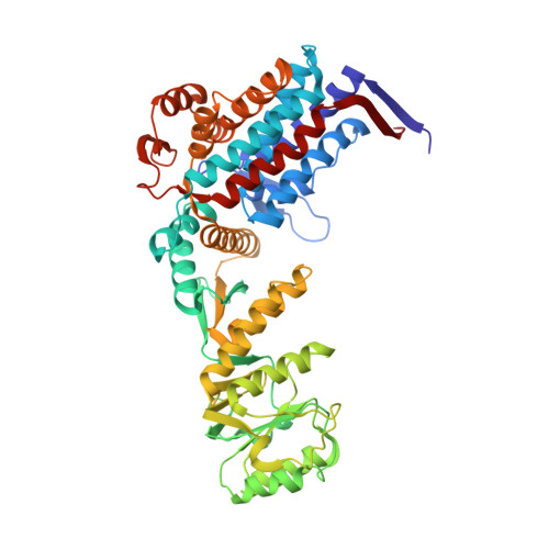
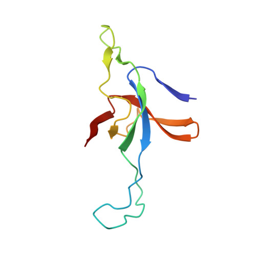
Crystal structure of groEL-groES


Experimental Data Snapshot

  • Method: X-RAY DIFFRACTION
  • Resolution: 2.81 Å
  • R-Value Free: 0.278 
  • R-Value Work: 0.262 

wwPDB Validation   3D Report Full Report


Ligand Structure Quality Assessment 


This is version 1.3 of the entry. See complete history


Literature

Role of the gamma-phosphate of ATP in triggering protein folding by GroEL-GroES: function, structure and energetics.

Chaudhry, C.Farr, G.W.Todd, M.J.Rye, H.S.Brunger, A.T.Adams, P.D.Horwich, A.L.Sigler, P.B.

(2003) EMBO J 22: 4877-4887

  • DOI: https://doi.org/10.1093/emboj/cdg477
  • Primary Citation of Related Structures:  
    1PCQ, 1PF9

  • PubMed Abstract: 

    Productive cis folding by the chaperonin GroEL is triggered by the binding of ATP but not ADP, along with cochaperonin GroES, to the same ring as non-native polypeptide, ejecting polypeptide into an encapsulated hydrophilic chamber. We examined the specific contribution of the gamma-phosphate of ATP to this activation process using complexes of ADP and aluminium or beryllium fluoride. These ATP analogues supported productive cis folding of the substrate protein, rhodanese, even when added to already-formed, folding-inactive cis ADP ternary complexes, essentially introducing the gamma-phosphate of ATP in an independent step. Aluminium fluoride was observed to stabilize the association of GroES with GroEL, with a substantial release of free energy (-46 kcal/mol). To understand the basis of such activation and stabilization, a crystal structure of GroEL-GroES-ADP.AlF3 was determined at 2.8 A. A trigonal AlF3 metal complex was observed in the gamma-phosphate position of the nucleotide pocket of the cis ring. Surprisingly, when this structure was compared with that of the previously determined GroEL-GroES-ADP complex, no other differences were observed. We discuss the likely basis of the ability of gamma-phosphate binding to convert preformed GroEL-GroES-ADP-polypeptide complexes into the folding-active state.


  • Organizational Affiliation

    Department of Molecular Biophysics and Biochemistry and Howard Hughes Medical Institute, Yale University, New Haven, CT, USA.


Macromolecules
Find similar proteins by:  (by identity cutoff)  |  3D Structure
Entity ID: 1
MoleculeChains Sequence LengthOrganismDetailsImage
groEL protein
A, B, C, D, E
A, B, C, D, E, F, G, H, I, J, K, L, M, N
524Escherichia coliMutation(s): 0 
Gene Names: GROL OR GROEL OR MOPA OR B4143 OR C5227 OR Z5748 OR ECS5124 OR SF4297
EC: 5.6.1.7
UniProt
Find proteins for P0A6F5 (Escherichia coli (strain K12))
Explore P0A6F5 
Go to UniProtKB:  P0A6F5
Entity Groups  
Sequence Clusters30% Identity50% Identity70% Identity90% Identity95% Identity100% Identity
UniProt GroupP0A6F5
Sequence Annotations
Expand
  • Reference Sequence
Find similar proteins by:  (by identity cutoff)  |  3D Structure
Entity ID: 2
MoleculeChains Sequence LengthOrganismDetailsImage
groES protein
O, P, Q, R, S
O, P, Q, R, S, T, U
97Escherichia coliMutation(s): 0 
Gene Names: GROS OR GROES OR MOPB OR B4142 OR C5226 OR Z5747 OR ECS5123 OR SF4296
UniProt
Find proteins for P0A6F9 (Escherichia coli (strain K12))
Explore P0A6F9 
Go to UniProtKB:  P0A6F9
Entity Groups  
Sequence Clusters30% Identity50% Identity70% Identity90% Identity95% Identity100% Identity
UniProt GroupP0A6F9
Sequence Annotations
Expand
  • Reference Sequence
Small Molecules
Ligands 4 Unique
IDChains Name / Formula / InChI Key2D Diagram3D Interactions
ADP
Query on ADP

Download Ideal Coordinates CCD File 
BA [auth B]
FA [auth C]
JA [auth D]
NA [auth E]
RA [auth F]
BA [auth B],
FA [auth C],
JA [auth D],
NA [auth E],
RA [auth F],
VA [auth G],
X [auth A]
ADENOSINE-5'-DIPHOSPHATE
C10 H15 N5 O10 P2
XTWYTFMLZFPYCI-KQYNXXCUSA-N
AF3
Query on AF3

Download Ideal Coordinates CCD File 
CA [auth B]
GA [auth C]
KA [auth D]
OA [auth E]
SA [auth F]
CA [auth B],
GA [auth C],
KA [auth D],
OA [auth E],
SA [auth F],
WA [auth G],
Y [auth A]
ALUMINUM FLUORIDE
Al F3
KLZUFWVZNOTSEM-UHFFFAOYSA-K
K
Query on K

Download Ideal Coordinates CCD File 
AA [auth B]
EA [auth C]
IA [auth D]
MA [auth E]
QA [auth F]
AA [auth B],
EA [auth C],
IA [auth D],
MA [auth E],
QA [auth F],
UA [auth G],
W [auth A]
POTASSIUM ION
K
NPYPAHLBTDXSSS-UHFFFAOYSA-N
MG
Query on MG

Download Ideal Coordinates CCD File 
DA [auth C]
HA [auth D]
LA [auth E]
PA [auth F]
TA [auth G]
DA [auth C],
HA [auth D],
LA [auth E],
PA [auth F],
TA [auth G],
V [auth A],
Z [auth B]
MAGNESIUM ION
Mg
JLVVSXFLKOJNIY-UHFFFAOYSA-N
Experimental Data & Validation

Experimental Data

  • Method: X-RAY DIFFRACTION
  • Resolution: 2.81 Å
  • R-Value Free: 0.278 
  • R-Value Work: 0.262 
  • Space Group: P 21 21 2
Unit Cell:
Length ( Å )Angle ( ˚ )
a = 255.546α = 90
b = 266.855β = 90
c = 187.049γ = 90
Software Package:
Software NamePurpose
REFMACrefinement
DENZOdata reduction
SCALEPACKdata scaling

Structure Validation

View Full Validation Report


Ligand Structure Quality Assessment 


Entry History 

Deposition Data

Revision History  (Full details and data files)

  • Version 1.0: 2003-10-14
    Type: Initial release
  • Version 1.1: 2008-04-29
    Changes: Version format compliance
  • Version 1.2: 2011-07-13
    Changes: Version format compliance
  • Version 1.3: 2024-02-14
    Changes: Data collection, Database references, Derived calculations, Refinement description