1PDG | pdb_00001pdg

CRYSTAL STRUCTURE OF HUMAN PLATELET-DERIVED GROWTH FACTOR BB


Experimental Data Snapshot

  • Method: X-RAY DIFFRACTION
  • Resolution: 3.00 Å
  • R-Value Observed: 
    0.209 (Depositor) 

wwPDB Validation 3D Report Full Report

Validation slider image for 1PDG

This is version 1.4 of the entry. See complete history

Literature

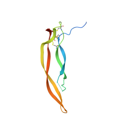
Crystal structure of human platelet-derived growth factor BB.

Oefner, C.D'Arcy, A.Winkler, F.K.Eggimann, B.Hosang, M.

(1992) EMBO J 11: 3921-3926

  • DOI: https://doi.org/10.1002/j.1460-2075.1992.tb05485.x
  • Primary Citation Related Structures: 
    1PDG

  • PubMed Abstract: 

    The crystal structure of the homodimeric BB isoform of human recombinant platelet-derived growth factor (PDGF-BB) has been determined by X-ray analysis to 3.0 A resolution. The polypeptide chain is folded into two highly twisted antiparallel pairs of beta-strands and contains an unusual knotted arrangement of three intramolecular disulfide bonds. Dimerization leads to the clustering of three surface loops at each end of the elongated dimer, which most probably form the receptor recognition sites.


  • Organizational Affiliation
    • Department of Pharmaceutical Research--New Technologies, F. Hoffmann-La Roche Ltd., Basel, Switzerland.

Macromolecule Content 

  • Total Structure Weight: 36.94 kDa 
  • Atom Count: 2,035 
  • Modeled Residue Count: 256 
  • Deposited Residue Count: 327 
  • Unique protein chains: 1

Macromolecules

Find similar proteins by:|  3D Structure
Entity ID: 1
MoleculeChains  Sequence LengthOrganismDetailsImage
PLATELET-DERIVED GROWTH FACTOR BB
A, B, C
109Homo sapiensMutation(s): 0 
UniProt & NIH Common Fund Data Resources
Find proteins for P01127 (Homo sapiens)
Explore P01127 
Go to UniProtKB:  P01127
PHAROS:  P01127
GTEx:  ENSG00000100311 
Entity Groups
Sequence Clusters30% Identity50% Identity70% Identity90% Identity95% Identity100% Identity
UniProt GroupP01127
Sequence Annotations
Expand
Reference Sequence

Experimental Data & Validation

Experimental Data

  • Method: X-RAY DIFFRACTION
  • Resolution: 3.00 Å
  • R-Value Observed: 0.209 (Depositor) 
Space Group: C 1 2 1
Unit Cell:
Length ( Å )Angle ( ˚ )
a = 147.3α = 90
b = 31.8β = 98.7
c = 90.1γ = 90
Software Package:
Software NamePurpose
PROLSQrefinement

Structure Validation

View Full Validation Report


Entry History 

Deposition Data

Revision History  (Full details and data files)

  • Version 1.0: 1994-01-31
    Type: Initial release
  • Version 1.1: 2008-03-24
    Changes: Version format compliance
  • Version 1.2: 2011-07-13
    Changes: Version format compliance
  • Version 1.3: 2017-11-29
    Changes: Derived calculations, Other
  • Version 1.4: 2024-11-13
    Changes: Data collection, Database references, Structure summary