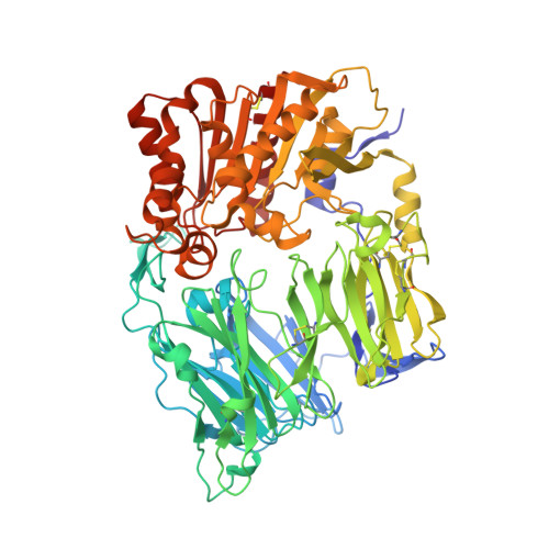
1PFQ

crystal structure of human apo dipeptidyl peptidase IV / CD26


Experimental Data Snapshot

  • Method: X-RAY DIFFRACTION
  • Resolution: 1.90 Å
  • R-Value Free: 0.298 
  • R-Value Work: 0.254 
  • R-Value Observed: 0.256 

wwPDB Validation   3D Report Full Report


Ligand Structure Quality Assessment 


This is version 1.3 of the entry. See complete history


Literature

High-resolution structure of human apo dipeptidyl peptidase IV/CD26 and its complex with 1-[([2-[(5-iodopyridin-2-yl)amino]-ethyl]amino)-acetyl]-2-cyano-(S)-pyrrolidine.

Oefner, C.D'Arcy, A.Mac Sweeney, A.Pierau, S.Gardiner, R.Dale, G.E.

(2003) Acta Crystallogr D Biol Crystallogr 59: 1206-1212

  • DOI: https://doi.org/10.1107/s0907444903010059
  • Primary Citation of Related Structures:  
    1PFQ

  • PubMed Abstract: 

    Dipeptidyl peptidase IV is a multifunctional type II transmembrane serine protease glycoprotein. The high-resolution crystal structure of the homodimeric human apo dipeptidyl peptidase IV has been determined at 1.9 A resolution. In addition, the structure of the binary complex with 1-[([2-[(5-iodopyridin-2-yl)amino]-ethyl]amino)-acetyl]-2-cyano-(S)-pyrrolidine has been solved, revealing the nature of the covalent interaction with the active-site serine.


  • Organizational Affiliation

    Morphochem AG, Basel, Switzerland. christian.oefner@morphochem.ch


Macromolecules
Find similar proteins by:  (by identity cutoff)  |  3D Structure
Entity ID: 1
MoleculeChains Sequence LengthOrganismDetailsImage
Dipeptidyl peptidase IV soluble form
A, B
731Homo sapiensMutation(s): 0 
Gene Names: DPP4 OR ADCP2 OR CD26
EC: 3.4.14.5
UniProt & NIH Common Fund Data Resources
Find proteins for P27487 (Homo sapiens)
Explore P27487 
Go to UniProtKB:  P27487
PHAROS:  P27487
GTEx:  ENSG00000197635 
Entity Groups  
Sequence Clusters30% Identity50% Identity70% Identity90% Identity95% Identity100% Identity
UniProt GroupP27487
Glycosylation
Glycosylation Sites: 2Go to GlyGen: P27487-1
Sequence Annotations
Expand
  • Reference Sequence
Experimental Data & Validation

Experimental Data

  • Method: X-RAY DIFFRACTION
  • Resolution: 1.90 Å
  • R-Value Free: 0.298 
  • R-Value Work: 0.254 
  • R-Value Observed: 0.256 
  • Space Group: P 21 21 21
Unit Cell:
Length ( Å )Angle ( ˚ )
a = 71.031α = 90
b = 118.142β = 90
c = 184.583γ = 90
Software Package:
Software NamePurpose
REFMACrefinement

Structure Validation

View Full Validation Report


Ligand Structure Quality Assessment 


Entry History 

Deposition Data

Revision History  (Full details and data files)

  • Version 1.0: 2003-07-01
    Type: Initial release
  • Version 1.1: 2008-04-29
    Changes: Version format compliance
  • Version 1.2: 2011-07-13
    Changes: Non-polymer description, Version format compliance
  • Version 1.3: 2020-07-29
    Type: Remediation
    Reason: Carbohydrate remediation
    Changes: Data collection, Derived calculations, Structure summary