1PHT | pdb_00001pht

PHOSPHATIDYLINOSITOL 3-KINASE P85-ALPHA SUBUNIT SH3 DOMAIN, RESIDUES 1-85


Experimental Data Snapshot

  • Method: X-RAY DIFFRACTION
  • Resolution: 2.00 Å
  • R-Value Free: 
    0.258 (Depositor) 
  • R-Value Work: 
    0.223 (Depositor) 
  • R-Value Observed: 
    0.223 (Depositor) 

wwPDB Validation 3D Report Full Report

Validation slider image for 1PHT

This is version 1.3 of the entry. See complete history

Literature

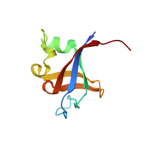
Crystal structure of P13K SH3 domain at 20 angstroms resolution.

Liang, J.Chen, J.K.Schreiber, S.T.Clardy, J.

(1996) J Mol Biology 257: 632-643

  • DOI: https://doi.org/10.1006/jmbi.1996.0190
  • Primary Citation Related Structures: 
    1PHT

  • PubMed Abstract: 

    The P13K SH3 domain, residues 1 to 85 of the P1-3 kinase p85 subunit, has been characterized by X-ray diffraction. Crystals belonging to space group P4(3)2(1)2 diffract to 2.0 angstroms resolution and the structure was phased by single isomorphous replacement and anomalous scattering (SIRAS). As expected, the domain is a compact beta barrel with an over-all confirmation very similar to the independently determined NMR structures. The X-ray structure illuminates a discrepancy between the two NMR structures on the conformation of the loop region unique to P13K SH3. Furthermore, the ligand binding pockets of P13K SH3 domain are occupied by amino acid residues from symmetry-related P13K SH3 molecules: the C-terminal residues I(82) SPP of one and R18 of another. The interaction modes clearly resemble those observed for the P13K SH3 domain complexed with the synthetic peptide RLP1, a class 1 ligand, although there are significant differences. The solid-state interactions suggest a model of protein-protein aggregation that could be mediated by SH3 domains.


  • Organizational Affiliation
    • Department of Chemistry, Baker Laboratory. Cornell University, Ithaca, NY 14853-1301, USA.

Macromolecule Content 

  • Total Structure Weight: 9.63 kDa 
  • Atom Count: 725 
  • Modeled Residue Count: 83 
  • Deposited Residue Count: 85 
  • Unique protein chains: 1

Macromolecules

Find similar proteins by:|  3D Structure
Entity ID: 1
MoleculeChains  Sequence LengthOrganismDetailsImage
PHOSPHATIDYLINOSITOL 3-KINASE P85-ALPHA SUBUNIT85Homo sapiensMutation(s): 0 
Gene Names: PI3K SH3 FROM CDNA LIBRARY OF
UniProt & NIH Common Fund Data Resources
Find proteins for P27986 (Homo sapiens)
Explore P27986 
Go to UniProtKB:  P27986
PHAROS:  P27986
GTEx:  ENSG00000145675 
Entity Groups
Sequence Clusters30% Identity50% Identity70% Identity90% Identity95% Identity100% Identity
UniProt GroupP27986
Sequence Annotations
Expand
Reference Sequence

Experimental Data & Validation

Experimental Data

  • Method: X-RAY DIFFRACTION
  • Resolution: 2.00 Å
  • R-Value Free:  0.258 (Depositor) 
  • R-Value Work:  0.223 (Depositor) 
  • R-Value Observed: 0.223 (Depositor) 
Space Group: P 43 21 2
Unit Cell:
Length ( Å )Angle ( ˚ )
a = 47α = 90
b = 47β = 90
c = 92.3γ = 90
Software Package:
Software NamePurpose
X-PLORmodel building
X-PLORrefinement
DENZOdata reduction
X-PLORphasing

Structure Validation

View Full Validation Report


Entry History 

Deposition Data

Revision History  (Full details and data files)

  • Version 1.0: 1995-12-07
    Type: Initial release
  • Version 1.1: 2008-03-24
    Changes: Version format compliance
  • Version 1.2: 2011-07-13
    Changes: Version format compliance
  • Version 1.3: 2024-02-14
    Changes: Data collection, Database references, Other