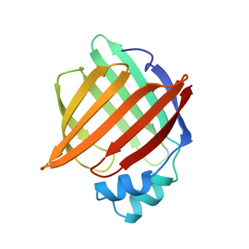
1PMP

CRYSTALLOGRAPHIC STUDIES ON A FAMILY OF CELLULAR LIPOPHILIC TRANSPORT PROTEINS. REFINEMENT OF P2 MYELIN PROTEIN AND THE STRUCTURE DETERMINATION AND REFINEMENT OF CELLULAR RETINOL-BINDING PROTEIN IN COMPLEX WITH ALL-TRANS-RETINOL


Experimental Data Snapshot

  • Method: X-RAY DIFFRACTION
  • Resolution: 2.70 Å
  • R-Value Work: 0.268 
  • R-Value Observed: 0.268 

wwPDB Validation   3D Report Full Report


This is version 1.3 of the entry. See complete history


Literature

Crystallographic studies on a family of cellular lipophilic transport proteins. Refinement of P2 myelin protein and the structure determination and refinement of cellular retinol-binding protein in complex with all-trans-retinol.

Cowan, S.W.Newcomer, M.E.Jones, T.A.

(1993) J Mol Biol 230: 1225-1246

  • DOI: https://doi.org/10.1006/jmbi.1993.1238
  • Primary Citation of Related Structures:  
    1CRB, 1PMP

  • PubMed Abstract: 

    P2 myelin protein (P2) and cellular retinol binding protein (CRBP) are members of a family of cellular lipophilic transport proteins. P2 has been refined at a resolution of 2.7 A, and CRBP has been solved by molecular replacement and refined to a resolution of 2.1 A. The members of this family form a compact three-dimensional structure built up from ten antiparallel strands that fold to form an orthogonal barrel containing the ligand. In P2, the carboxylate group of an oleic acid ligand interacts with the side-chains of two arginine (106 and 126), and one tyrosine (128) residues. The ligand adopts a U-shaped conformation. In CRBP, the all-trans-retinol has a planar conformation with its alcohol group hydrogen bonding to the side-chain of glutamine 108 (equivalent to residue 106 in P2). The local interactions of glutamine 108 explain CRBP's preference for binding retinol rather than retinal. The side-chain of lysine 40 makes a close contact with the isoprene tail of the retinol.


  • Organizational Affiliation

    Department of Molecular Biology, Uppsala University Biomedical Centre, Sweden.


Macromolecules
Find similar proteins by:  (by identity cutoff)  |  3D Structure
Entity ID: 1
MoleculeChains Sequence LengthOrganismDetailsImage
P2 MYELIN PROTEIN
A, B, C
131Bos taurusMutation(s): 0 
UniProt
Find proteins for P02690 (Bos taurus)
Explore P02690 
Go to UniProtKB:  P02690
Entity Groups  
Sequence Clusters30% Identity50% Identity70% Identity90% Identity95% Identity100% Identity
UniProt GroupP02690
Sequence Annotations
Expand
  • Reference Sequence
Experimental Data & Validation

Experimental Data

  • Method: X-RAY DIFFRACTION
  • Resolution: 2.70 Å
  • R-Value Work: 0.268 
  • R-Value Observed: 0.268 
  • Space Group: P 21 21 21
Unit Cell:
Length ( Å )Angle ( ˚ )
a = 91.8α = 90
b = 99.5β = 90
c = 56.5γ = 90
Software Package:
Software NamePurpose
X-PLORmodel building
X-PLORrefinement
X-PLORphasing

Structure Validation

View Full Validation Report


Entry History 

Deposition Data

Revision History  (Full details and data files)

  • Version 1.0: 1995-01-26
    Type: Initial release
  • Version 1.1: 2008-03-24
    Changes: Version format compliance
  • Version 1.2: 2011-07-13
    Changes: Version format compliance
  • Version 1.3: 2024-02-14
    Changes: Data collection, Database references, Derived calculations, Other