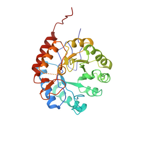
1Q3N

Crystal structure of KDO8P synthase in its binary complex with substrate PEP


Experimental Data Snapshot

  • Method: X-RAY DIFFRACTION
  • Resolution: 2.70 Å
  • R-Value Free: 0.268 
  • R-Value Work: 0.235 
  • R-Value Observed: 0.242 

Starting Model: experimental
View more details

wwPDB Validation   3D Report Full Report


Ligand Structure Quality Assessment 


This is version 1.3 of the entry. See complete history


Literature

Crystal structures of Escherichia coli KDO8P synthase complexes reveal the source of catalytic irreversibility.

Vainer, R.Belakhov, V.Rabkin, E.Baasov, T.Adir, N.

(2005) J Mol Biol 351: 641-652

  • DOI: https://doi.org/10.1016/j.jmb.2005.06.021
  • Primary Citation of Related Structures:  
    1PHW, 1Q3N, 1X6U, 1X8F

  • PubMed Abstract: 

    The enzyme 3-deoxy-D-manno-2-octulosonate-8-phosphate synthase (KDO8PS) catalyses the condensation of arabinose 5-phosphate (A5P) and phosphoenol pyruvate (PEP) to obtain 3-deoxy-D-manno-2-octulosonate-8-phosphate (KDO8P). We have elucidated initial modes of ligand binding in KDO8PS binary complexes by X-ray crystallography. Structures of the apo-enzyme and of binary complexes with the substrate PEP, the product KDO8P and the catalytically inactive 1-deoxy analog of arabinose 5-phosphate (1dA5P) were obtained. The KDO8PS active site resembles an irregular funnel with positive electrostatic potential situated at the bottom of the PEP-binding sub-site, which is the primary attractive force towards negatively charged phosphate moieties of all ligands. The structures of the ligand-free apo-KDO8PS and the binary complex with the product KDO8P visualize for the first time the role of His202 as an active-site gate. Examination of the crystal structures of KDO8PS with the KDO8P or 1dA5P shows these ligands bound to the enzyme in the PEP-binding sub-site, and not as expected to the A5P sub-site. Taken together, the structures presented here strengthen earlier evidence that this enzyme functions predominantly through positional catalysis, map out the roles of active-site residues and provide evidence that explains the total lack of catalytic reversibility.


  • Organizational Affiliation

    Department of Chemistry and Institute of Catalysis, Science and Technology, Technion-Israel Institute of Technology, Technion City, Haifa 32000, Israel.


Macromolecules
Find similar proteins by:  (by identity cutoff)  |  3D Structure
Entity ID: 1
MoleculeChains Sequence LengthOrganismDetailsImage
2-dehydro-3-deoxyphosphooctonate aldolase284Escherichia coliMutation(s): 0 
Gene Names: KDSA
EC: 2.5.1.55
UniProt
Find proteins for P0A715 (Escherichia coli (strain K12))
Explore P0A715 
Go to UniProtKB:  P0A715
Entity Groups  
Sequence Clusters30% Identity50% Identity70% Identity90% Identity95% Identity100% Identity
UniProt GroupP0A715
Sequence Annotations
Expand
  • Reference Sequence
Small Molecules
Ligands 1 Unique
IDChains Name / Formula / InChI Key2D Diagram3D Interactions
PEP
Query on PEP

Download Ideal Coordinates CCD File 
B [auth A]PHOSPHOENOLPYRUVATE
C3 H5 O6 P
DTBNBXWJWCWCIK-UHFFFAOYSA-N
Experimental Data & Validation

Experimental Data

  • Method: X-RAY DIFFRACTION
  • Resolution: 2.70 Å
  • R-Value Free: 0.268 
  • R-Value Work: 0.235 
  • R-Value Observed: 0.242 
  • Space Group: I 2 3
Unit Cell:
Length ( Å )Angle ( ˚ )
a = 118.441α = 90
b = 118.441β = 90
c = 118.441γ = 90
Software Package:
Software NamePurpose
DENZOdata reduction
SCALEPACKdata scaling
CNSrefinement
CNSphasing

Structure Validation

View Full Validation Report


Ligand Structure Quality Assessment 


Entry History 

Deposition Data

Revision History  (Full details and data files)

  • Version 1.0: 2004-10-12
    Type: Initial release
  • Version 1.1: 2008-04-29
    Changes: Version format compliance
  • Version 1.2: 2011-07-13
    Changes: Derived calculations, Version format compliance
  • Version 1.3: 2023-08-16
    Changes: Data collection, Database references, Derived calculations, Refinement description