1Q7G | pdb_00001q7g

Homoserine Dehydrogenase in complex with suicide inhibitor complex NAD-5-hydroxy-4-Oxonorvaline


Experimental Data Snapshot

  • Method: X-RAY DIFFRACTION
  • Resolution: 2.60 Å
  • R-Value Free: 
    0.306 (Depositor), 0.297 (DCC) 
  • R-Value Work: 
    0.247 (Depositor), 0.234 (DCC) 
  • R-Value Observed: 
    0.247 (Depositor) 

Starting Model: experimental
View more details

wwPDB Validation 3D Report Full Report

Validation slider image for 1Q7G

Ligand Structure Quality Assessment 


This is version 1.3 of the entry. See complete history

Literature

Enzyme assisted suicide: Molecular basis for the antifungal activity of 5-hydroxy-4-oxonorvaline by potent inhibition of homoserine dehydrogenase

Jacques, S.L.Mirza, I.A.Ejim, L.Koteva, K.Hughes, D.W.Green, K.Kinach, R.Honek, J.F.Lai, H.K.Berghuis, A.M.Wright, G.D.

(2003) Chem Biol 10: 989-995

  • DOI: https://doi.org/10.1016/j.chembiol.2003.09.015
  • Primary Citation Related Structures: 
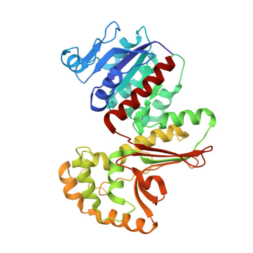
    1Q7G

  • PubMed Abstract: 

    The structure of the antifungal drug 5-hydroxy-4-oxonorvaline (HON) in complex with its target homoserine dehydrogenase (HSD) has been determined by X-ray diffraction to 2.6 A resolution. HON shows potent in vitro and in vivo activity against various fungal pathogens despite its weak (2 mM) affinity for HSD in the steady state. The structure together with structure-activity relationship studies, mass spectrometry experiments, and spectroscopic data reveals that the molecular mechanism of antifungal action conferred by HON involves enzyme-dependent formation of a covalent adduct between C4 of the nicotinamide ring of NAD(+) and C5 of HON. Furthermore, novel interactions are involved in stabilizing the (HON*NAD)-adduct, which are not observed in the enzyme's ternary complex structure. These findings clarify the apparent paradox of the potent antifungal actions of HON given its weak steady-state inhibition characteristics.


  • Organizational Affiliation
    • Antimicrobial Research Centre and Department of Biochemistry, McMaster University, Hamilton L8N 3Z5, Canada.

Macromolecule Content 

  • Total Structure Weight: 77.95 kDa 
  • Atom Count: 5,571 
  • Modeled Residue Count: 716 
  • Deposited Residue Count: 718 
  • Unique protein chains: 1

Macromolecules

Find similar proteins by:|  3D Structure
Entity ID: 1
MoleculeChains  Sequence LengthOrganismDetailsImage
Homoserine dehydrogenase
A, B
359Saccharomyces cerevisiaeMutation(s): 0 
Gene Names: HOM6YJR139C OR J2132
EC: 1.1.1.3
UniProt
Find proteins for P31116 (Saccharomyces cerevisiae (strain ATCC 204508 / S288c))
Explore P31116 
Go to UniProtKB:  P31116
Entity Groups
Sequence Clusters30% Identity50% Identity70% Identity90% Identity95% Identity100% Identity
UniProt GroupP31116
Sequence Annotations
Expand
Reference Sequence

Experimental Data & Validation

Experimental Data

  • Method: X-RAY DIFFRACTION
  • Resolution: 2.60 Å
  • R-Value Free:  0.306 (Depositor), 0.297 (DCC) 
  • R-Value Work:  0.247 (Depositor), 0.234 (DCC) 
  • R-Value Observed: 0.247 (Depositor) 
Space Group: P 43 21 2
Unit Cell:
Length ( Å )Angle ( ˚ )
a = 80.863α = 90
b = 80.863β = 90
c = 248.986γ = 90
Software Package:
Software NamePurpose
CNSrefinement
SCALEPACKdata scaling
CNSphasing

Structure Validation

View Full Validation Report


Ligand Structure Quality Assessment 


Entry History 

Deposition Data

Revision History  (Full details and data files)

  • Version 1.0: 2003-10-21
    Type: Initial release
  • Version 1.1: 2008-04-29
    Changes: Version format compliance
  • Version 1.2: 2011-07-13
    Changes: Version format compliance
  • Version 1.3: 2023-08-16
    Changes: Data collection, Database references, Derived calculations, Refinement description