1QMT

Recombinant Human Eosinophil Cationic Protein


Experimental Data Snapshot

  • Method: X-RAY DIFFRACTION
  • Resolution: 2.40 Å
  • R-Value Free: 0.229 
  • R-Value Work: 0.176 
  • R-Value Observed: 0.176 

wwPDB Validation   3D Report Full Report


This is version 1.2 of the entry. See complete history


Literature

The Crystal Structure of Eosinophil Cationic Protein at 2.4 A Resolution

Boix, E.Leonidas, D.D.Nikolovski, Z.Nogues, M.V.Cuchillo, C.M.Acharya, K.R.

(1999) Biochemistry 38: 16794

  • DOI: https://doi.org/10.1021/bi9919145
  • Primary Citation of Related Structures:  
    1QMT

  • PubMed Abstract: 

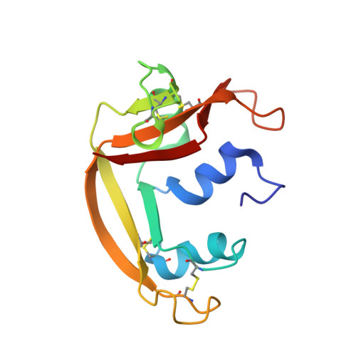
    Eosinophil cationic protein (ECP) is located in the matrix of the eosinophil's large specific granule and has marked toxicity for a variety of helminth parasites, hemoflagellates, bacteria, single-stranded RNA virus, and mammalian cells and tissues. It belongs to the bovine pancreatic ribonuclease A (RNase A) family and exhibits ribonucleolytic activity which is about 100-fold lower than that of a related eosinophil ribonuclease, the eosinophil-derived neurotoxin (EDN). The crystal structure of human ECP, determined at 2.4 A, is similar to that of RNase A and EDN. It reveals that residues Gln-14, His-15, Lys-38, Thr-42, and His-128 at the active site are conserved as in all other RNase A homologues. Nevertheless, evidence for considerable divergence of ECP is also implicit in the structure. Amino acid residues Arg-7, Trp-10, Asn-39, His-64, and His-82 appear to play a key part in the substrate specificity and low catalytic activity of ECP. The structure also shows how the cationic residues are distributed on the surface of the ECP molecule that may have implications for an understanding of the cytotoxicity of this enzyme.


  • Organizational Affiliation

    Department of Biology and Biochemistry, University of Bath, UK.


Macromolecules
Find similar proteins by:  (by identity cutoff)  |  3D Structure
Entity ID: 1
MoleculeChains Sequence LengthOrganismDetailsImage
EOSINOPHIL CATIONIC PROTEIN134Homo sapiensMutation(s): 0 
UniProt & NIH Common Fund Data Resources
Find proteins for P12724 (Homo sapiens)
Explore P12724 
Go to UniProtKB:  P12724
PHAROS:  P12724
GTEx:  ENSG00000169397 
Entity Groups  
Sequence Clusters30% Identity50% Identity70% Identity90% Identity95% Identity100% Identity
UniProt GroupP12724
Sequence Annotations
Expand
  • Reference Sequence
Experimental Data & Validation

Experimental Data

  • Method: X-RAY DIFFRACTION
  • Resolution: 2.40 Å
  • R-Value Free: 0.229 
  • R-Value Work: 0.176 
  • R-Value Observed: 0.176 
  • Space Group: P 63
Unit Cell:
Length ( Å )Angle ( ˚ )
a = 100.159α = 90
b = 100.159β = 90
c = 31.281γ = 120
Software Package:
Software NamePurpose
X-PLORrefinement
DENZOdata reduction
SCALEPACKdata scaling
X-PLORphasing

Structure Validation

View Full Validation Report


Entry History 

Deposition Data

Revision History  (Full details and data files)

  • Version 1.0: 2000-02-04
    Type: Initial release
  • Version 1.1: 2011-05-08
    Changes: Version format compliance
  • Version 1.2: 2011-07-13
    Changes: Version format compliance