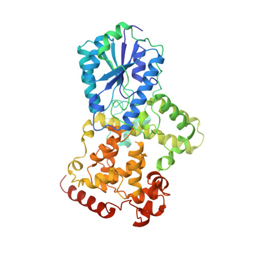
1QNF

STRUCTURE OF PHOTOLYASE


Experimental Data Snapshot

  • Method: X-RAY DIFFRACTION
  • Resolution: 1.80 Å
  • R-Value Free: 0.239 
  • R-Value Work: 0.197 
  • R-Value Observed: 0.197 

wwPDB Validation   3D Report Full Report


Ligand Structure Quality Assessment 


This is version 1.3 of the entry. See complete history


Literature

Crystal structure of DNA photolyase from Anacystis nidulans

Tamada, T.Kitadokoro, K.Higuchi, Y.Inaka, K.Yasui, A.de Ruiter, P.E.Eker, A.P.Miki, K.

(1997) Nat Struct Biol 4: 887-891

  • DOI: https://doi.org/10.1038/nsb1197-887
  • Primary Citation of Related Structures:  
    1QNF

  • PubMed Abstract: 

    The crystal structure at 1.8 A resolution of 8-HDF type photolyase from A. nidulans shows a backbone structure similar to that of MTHF type E. coli photolyase but reveals a completely different binding site for the light-harvesting cofactor.


Macromolecules
Find similar proteins by:  (by identity cutoff)  |  3D Structure
Entity ID: 1
MoleculeChains Sequence LengthOrganismDetailsImage
PHOTOLYASE484Synechococcus elongatus PCC 6301Mutation(s): 0 
EC: 4.1.99.3
UniProt
Find proteins for P05327 (Synechococcus sp. (strain ATCC 27144 / PCC 6301 / SAUG 1402/1))
Explore P05327 
Go to UniProtKB:  P05327
Entity Groups  
Sequence Clusters30% Identity50% Identity70% Identity90% Identity95% Identity100% Identity
UniProt GroupP05327
Sequence Annotations
Expand
  • Reference Sequence
Small Molecules
Ligands 2 Unique
IDChains Name / Formula / InChI Key2D Diagram3D Interactions
FAD
Query on FAD

Download Ideal Coordinates CCD File 
B [auth A]FLAVIN-ADENINE DINUCLEOTIDE
C27 H33 N9 O15 P2
VWWQXMAJTJZDQX-UYBVJOGSSA-N
HDF
Query on HDF

Download Ideal Coordinates CCD File 
C [auth A]8-HYDROXY-10-(D-RIBO-2,3,4,5-TETRAHYDROXYPENTYL)-5-DEAZAISOALLOXAZINE
C16 H17 N3 O7
AUEILLWDYUBWCM-AGIUHOORSA-N
Experimental Data & Validation

Experimental Data

  • Method: X-RAY DIFFRACTION
  • Resolution: 1.80 Å
  • R-Value Free: 0.239 
  • R-Value Work: 0.197 
  • R-Value Observed: 0.197 
  • Space Group: P 43 21 2
Unit Cell:
Length ( Å )Angle ( ˚ )
a = 91.17α = 90
b = 91.17β = 90
c = 135.54γ = 90
Software Package:
Software NamePurpose
X-PLORmodel building
X-PLORrefinement
DENZOdata reduction
SCALEPACKdata scaling
X-PLORphasing

Structure Validation

View Full Validation Report


Ligand Structure Quality Assessment 


Entry History 

Deposition Data

Revision History  (Full details and data files)

  • Version 1.0: 1998-01-14
    Type: Initial release
  • Version 1.1: 2008-03-24
    Changes: Version format compliance
  • Version 1.2: 2011-07-13
    Changes: Source and taxonomy, Version format compliance
  • Version 1.3: 2024-02-14
    Changes: Data collection, Database references, Derived calculations, Other