1QRZ

CATALYTIC DOMAIN OF PLASMINOGEN


Experimental Data Snapshot

  • Method: X-RAY DIFFRACTION
  • Resolution: 2.00 Å
  • R-Value Free: 0.294 
  • R-Value Work: 0.235 
  • R-Value Observed: 0.235 

wwPDB Validation   3D Report Full Report


This is version 1.4 of the entry. See complete history


Literature

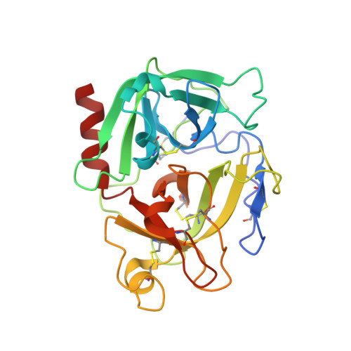
Crystal structure of the proenzyme domain of plasminogen.

Peisach, E.Wang, J.de los Santos, T.Reich, E.Ringe, D.

(1999) Biochemistry 38: 11180-11188

  • DOI: https://doi.org/10.1021/bi991130r
  • Primary Citation of Related Structures:  
    1QRZ

  • PubMed Abstract: 

    We have solved the X-ray crystal structure of the proenzyme form of the catalytic domain of plasminogen, with the nonessential mutations M585Q, V673M, and M788L, to 2.0 A resolution. The structure presents an inactive protease characterized by Asp740 (chymotrypsinogen 194) hydrogen bonded to His586 (chymotrypsinogen 40), preventing proper formation of the oxyanion hole and S1 specificity pocket. In addition, the catalytic triad residues are misplaced relative to the active conformation adopted by serine proteases in the chymotrypsin family. Finally, a unique form of zymogen inactivation is observed, characterized by a "foot-in-mouth" mechanism in which Trp761 (chymotrypsinogen 215) is folded into the S1 specificity pocket preventing substrate binding.


  • Organizational Affiliation

    Program in Biophysics and Structural Biology, Brandeis University, Waltham, Massachusetts 02454-9110, USA.


Macromolecules
Find similar proteins by:  (by identity cutoff)  |  3D Structure
Entity ID: 1
MoleculeChains Sequence LengthOrganismDetailsImage
PLASMINOGEN
A, B, C, D
246Homo sapiensMutation(s): 3 
UniProt & NIH Common Fund Data Resources
Find proteins for P00747 (Homo sapiens)
Explore P00747 
Go to UniProtKB:  P00747
PHAROS:  P00747
GTEx:  ENSG00000122194 
Entity Groups  
Sequence Clusters30% Identity50% Identity70% Identity90% Identity95% Identity100% Identity
UniProt GroupP00747
Sequence Annotations
Expand
  • Reference Sequence
Experimental Data & Validation

Experimental Data

  • Method: X-RAY DIFFRACTION
  • Resolution: 2.00 Å
  • R-Value Free: 0.294 
  • R-Value Work: 0.235 
  • R-Value Observed: 0.235 
  • Space Group: P 1 21 1
Unit Cell:
Length ( Å )Angle ( ˚ )
a = 81.658α = 90
b = 73.575β = 108.73
c = 81.363γ = 90
Software Package:
Software NamePurpose
DENZOdata reduction
SCALEPACKdata scaling
AMoREphasing
CNSrefinement

Structure Validation

View Full Validation Report


Entry History 

Deposition Data

Revision History  (Full details and data files)

  • Version 1.0: 1999-10-14
    Type: Initial release
  • Version 1.1: 2008-04-27
    Changes: Version format compliance
  • Version 1.2: 2011-07-13
    Changes: Version format compliance
  • Version 1.3: 2013-09-25
    Changes: Derived calculations
  • Version 1.4: 2021-11-03
    Changes: Database references