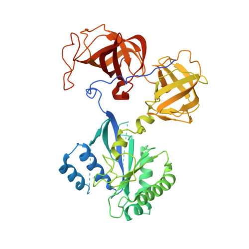
Crystal structure and functional analysis of the eukaryotic class II release factor eRF3 from S. pombe
Kong, C., Ito, K., Walsh, M.A., Wada, M., Liu, Y., Kumar, S., Barford, D., Nakamura, Y., Song, H.(2004) Mol Cell 14: 233-245
- PubMed: 15099522
- DOI: https://doi.org/10.1016/s1097-2765(04)00206-0
- Primary Citation of Related Structures:
1R5B, 1R5N, 1R5O - PubMed Abstract:
Translation termination in eukaryotes is governed by two interacting release factors, eRF1 and eRF3. The crystal structure of the eEF1alpha-like region of eRF3 from S. pombe determined in three states (free protein, GDP-, and GTP-bound forms) reveals an overall structure that is similar to EF-Tu, although with quite different domain arrangements. In contrast to EF-Tu, GDP/GTP binding to eRF3c does not induce dramatic conformational changes, and Mg(2+) is not required for GDP binding to eRF3c. Mg(2+) at higher concentration accelerates GDP release, suggesting a novel mechanism for nucleotide exchange on eRF3 from that of other GTPases. Mapping sequence conservation onto the molecular surface, combined with mutagenesis analysis, identified the eRF1 binding region, and revealed an essential function for the C terminus of eRF3. The N-terminal extension, rich in acidic amino acids, blocks the proposed eRF1 binding site, potentially regulating eRF1 binding to eRF3 in a competitive manner.
- Laboratory of Macromolecular Structure, Institute of Molecular and Cell Biology, 30 Medical Drive, Singapore 117609, Japan.
Organizational Affiliation:


