1RJB

Crystal Structure of FLT3


Experimental Data Snapshot

  • Method: X-RAY DIFFRACTION
  • Resolution: 2.10 Å
  • R-Value Free: 0.248 
  • R-Value Work: 0.210 
  • R-Value Observed: 0.220 

wwPDB Validation   3D Report Full Report


This is version 1.3 of the entry. See complete history


Literature

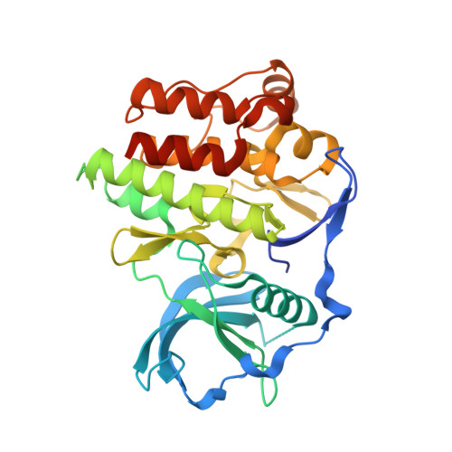
The structural basis for autoinhibition of FLT3 by the juxtamembrane domain.

Griffith, J.Black, J.Faerman, C.Swenson, L.Wynn, M.Lu, F.Lippke, J.Saxena, K.

(2004) Mol Cell 13: 169-178

  • DOI: https://doi.org/10.1016/s1097-2765(03)00505-7
  • Primary Citation of Related Structures:  
    1RJB

  • PubMed Abstract: 

    FLT3 is a type III receptor tyrosine kinase that is thought to play a key role in hematopoiesis. Certain classes of FLT3 mutations cause constitutively activated forms of the receptor that are found in significant numbers of patients with acute myelogenous leukemia (AML). The mutations occur either in the activation loop, for example, as point mutations of Asp835 or as internal tandem duplication (ITD) sequences in the juxtamembrane (JM) domain. To further understand the nature of FLT3 autoinhibition and regulation, we have determined the crystal structure of the autoinhibited form of FLT3. This structure shows the autoinhibitory conformation of a complete JM domain in this class of receptor tyrosine kinases. The detailed inhibitory mechanism of the JM domain is revealed, which is likely utilized by other members of type III receptor tyrosine kinases.


  • Organizational Affiliation

    Vertex Pharmaceuticals Incorporated, 130 Waverly Street, Cambridge, MA 02139, USA. james_griffith@vrtx.com


Macromolecules
Find similar proteins by:  (by identity cutoff)  |  3D Structure
Entity ID: 1
MoleculeChains Sequence LengthOrganismDetailsImage
FL cytokine receptor344Homo sapiensMutation(s): 0 
EC: 2.7.1.112
UniProt & NIH Common Fund Data Resources
Find proteins for P36888 (Homo sapiens)
Explore P36888 
Go to UniProtKB:  P36888
PHAROS:  P36888
GTEx:  ENSG00000122025 
Entity Groups  
Sequence Clusters30% Identity50% Identity70% Identity90% Identity95% Identity100% Identity
UniProt GroupP36888
Sequence Annotations
Expand
  • Reference Sequence
Experimental Data & Validation

Experimental Data

  • Method: X-RAY DIFFRACTION
  • Resolution: 2.10 Å
  • R-Value Free: 0.248 
  • R-Value Work: 0.210 
  • R-Value Observed: 0.220 
  • Space Group: P 43 21 2
Unit Cell:
Length ( Å )Angle ( ˚ )
a = 80.651α = 90
b = 80.651β = 90
c = 150.12γ = 90
Software Package:
Software NamePurpose
CNXrefinement
HKL-2000data reduction
CrystalCleardata scaling
AMoREphasing

Structure Validation

View Full Validation Report


Entry History 

Deposition Data

Revision History  (Full details and data files)

  • Version 1.0: 2004-02-03
    Type: Initial release
  • Version 1.1: 2008-04-29
    Changes: Version format compliance
  • Version 1.2: 2011-07-13
    Changes: Version format compliance
  • Version 1.3: 2024-02-14
    Changes: Data collection, Database references