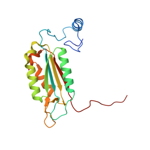
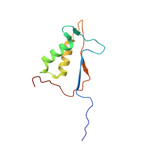
1RWP

Crystal structure of human caspase-1 in complex with 3-{6-[(8-hydroxy-quinoline-2-carbonyl)-amino]-2-thiophen-2-yl-hexanoylamino}-4-oxo-butyric acid


Experimental Data Snapshot

  • Method: X-RAY DIFFRACTION
  • Resolution: 2.20 Å
  • R-Value Free: 0.245 
  • R-Value Work: 0.212 
  • R-Value Observed: 0.214 

Starting Model: experimental
View more details

wwPDB Validation   3D Report Full Report


Ligand Structure Quality Assessment 


This is version 1.4 of the entry. See complete history


Literature

Structural analysis of caspase-1 inhibitors derived from Tethering.

O'Brien, T.Fahr, B.T.Sopko, M.M.Lam, J.W.Waal, N.D.Raimundo, B.C.Purkey, H.E.Pham, P.Romanowski, M.J.

(2005) Acta Crystallogr Sect F Struct Biol Cryst Commun 61: 451-458

  • DOI: https://doi.org/10.1107/S1744309105010109
  • Primary Citation of Related Structures:  
    1RWK, 1RWM, 1RWN, 1RWO, 1RWP

  • PubMed Abstract: 

    Caspase-1 is a key endopeptidase responsible for the post-translational processing of the IL-1beta and IL-18 cytokines and small-molecule inhibitors that modulate the activity of this enzyme are predicted to be important therapeutic treatments for many inflammatory diseases. A fragment-assembly approach, accompanied by structural analysis, was employed to generate caspase-1 inhibitors. With the aid of Tethering with extenders (small molecules that bind to the active-site cysteine and contain a free thiol), two novel fragments that bound to the active site and made a disulfide bond with the extender were identified by mass spectrometry. Direct linking of each fragment to the extender generated submicromolar reversible inhibitors that significantly reduced secretion of IL-1beta but not IL-6 from human peripheral blood mononuclear cells. Thus, Tethering with extenders facilitated rapid identification and synthesis of caspase-1 inhibitors with cell-based activity and subsequent structural analyses provided insights into the enzyme's ability to accommodate different inhibitor-binding modes in the active site.


  • Organizational Affiliation

    Department of Biology, Sunesis Pharmaceuticals Inc., USA.


Macromolecules
Find similar proteins by:  (by identity cutoff)  |  3D Structure
Entity ID: 1
MoleculeChains Sequence LengthOrganismDetailsImage
Interleukin-1 beta convertase178Homo sapiensMutation(s): 0 
Gene Names: CASP1IL1BCIL1BCE
EC: 3.4.22.36
UniProt & NIH Common Fund Data Resources
Find proteins for P29466 (Homo sapiens)
Explore P29466 
Go to UniProtKB:  P29466
PHAROS:  P29466
GTEx:  ENSG00000137752 
Entity Groups  
Sequence Clusters30% Identity50% Identity70% Identity90% Identity95% Identity100% Identity
UniProt GroupP29466
Sequence Annotations
Expand
  • Reference Sequence
Find similar proteins by:  (by identity cutoff)  |  3D Structure
Entity ID: 2
MoleculeChains Sequence LengthOrganismDetailsImage
Interleukin-1 beta convertase88Homo sapiensMutation(s): 0 
Gene Names: CASP1IL1BCIL1BCE
EC: 3.4.22.36
UniProt & NIH Common Fund Data Resources
Find proteins for P29466 (Homo sapiens)
Explore P29466 
Go to UniProtKB:  P29466
PHAROS:  P29466
GTEx:  ENSG00000137752 
Entity Groups  
Sequence Clusters30% Identity50% Identity70% Identity90% Identity95% Identity100% Identity
UniProt GroupP29466
Sequence Annotations
Expand
  • Reference Sequence
Small Molecules
Ligands 1 Unique
IDChains Name / Formula / InChI Key2D Diagram3D Interactions
HQC
Query on HQC

Download Ideal Coordinates CCD File 
C [auth A]3-{6-[(8-HYDROXY-QUINOLINE-2-CARBONYL)-AMINO]-2-THIOPHEN-2-YL-HEXANOYLAMINO}-4-OXO-BUTYRI ACID
C24 H25 N3 O6 S
QXMPYGCWDMKRMF-IRXDYDNUSA-N
Experimental Data & Validation

Experimental Data

  • Method: X-RAY DIFFRACTION
  • Resolution: 2.20 Å
  • R-Value Free: 0.245 
  • R-Value Work: 0.212 
  • R-Value Observed: 0.214 
  • Space Group: P 43 21 2
Unit Cell:
Length ( Å )Angle ( ˚ )
a = 63.216α = 90
b = 63.216β = 90
c = 161.694γ = 90
Software Package:
Software NamePurpose
REFMACrefinement
CrystalCleardata reduction
d*TREKdata scaling
AMoREphasing

Structure Validation

View Full Validation Report


Ligand Structure Quality Assessment 


Entry History 

Deposition Data

Revision History  (Full details and data files)

  • Version 1.0: 2004-12-28
    Type: Initial release
  • Version 1.1: 2008-04-29
    Changes: Version format compliance
  • Version 1.2: 2011-07-13
    Changes: Derived calculations, Version format compliance
  • Version 1.3: 2023-08-23
    Changes: Data collection, Database references, Derived calculations, Refinement description
  • Version 1.4: 2024-11-20
    Changes: Structure summary