1SR5 | pdb_00001sr5

ANTITHROMBIN-ANHYDROTHROMBIN-HEPARIN TERNARY COMPLEX STRUCTURE


Experimental Data Snapshot

  • Method: X-RAY DIFFRACTION
  • Resolution: 3.10 Å
  • R-Value Free: 
    0.278 (Depositor), 0.273 (DCC) 
  • R-Value Work: 
    0.227 (Depositor), 0.230 (DCC) 

Starting Models: experimental
View more details

wwPDB Validation 3D Report Full Report

Validation slider image for 1SR5

Ligand Structure Quality Assessment 


This is version 3.2 of the entry. See complete history

Literature

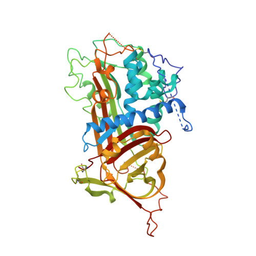
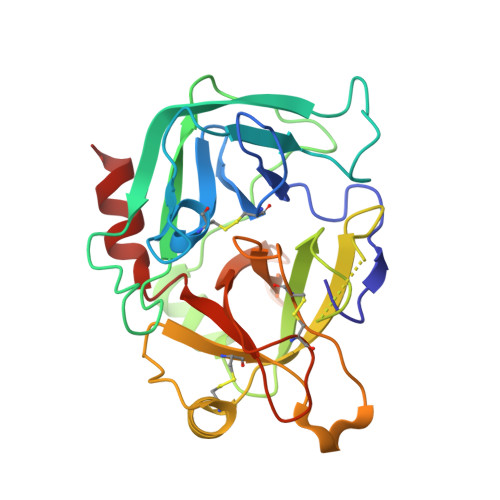
The ternary complex of antithrombin-anhydrothrombin-heparin reveals the basis of inhibitor specificity.

Dementiev, A.Petitou, M.Herbert, J.M.Gettins, P.G.

(2004) Nat Struct Mol Biol 11: 863-867

  • DOI: https://doi.org/10.1038/nsmb810
  • Primary Citation Related Structures: 
    1SR5

  • PubMed Abstract: 

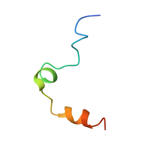
    Antithrombin, the principal physiological inhibitor of the blood coagulation proteinase thrombin, requires heparin as a cofactor. We report the crystal structure of the rate-determining encounter complex formed between antithrombin, anhydrothrombin and an optimal synthetic 16-mer oligosaccharide. The antithrombin reactive center loop projects from the serpin body and adopts a canonical conformation that makes extensive backbone and side chain contacts from P5 to P6' with thrombin's restrictive specificity pockets, including residues in the 60-loop. These contacts rationalize many earlier mutagenesis studies on thrombin specificity. The 16-mer oligosaccharide is just long enough to form the predicted bridge between the high-affinity pentasaccharide-binding site on antithrombin and the highly basic exosite 2 on thrombin, validating the design strategy for this synthetic heparin. The protein-protein and protein-oligosaccharide interactions together explain the basis for heparin activation of antithrombin as a thrombin inhibitor.


  • Organizational Affiliation
    • Department of Biochemistry and Molecular Genetics, College of Medicine, University of Illinois at Chicago, 900 S. Ashland, Chicago, Illinois 60607, USA.

Macromolecule Content 

  • Total Structure Weight: 85.96 kDa 
  • Atom Count: 5,534 
  • Modeled Residue Count: 689 
  • Deposited Residue Count: 727 
  • Unique protein chains: 3

Macromolecules

Find similar proteins by:|  3D Structure
Entity ID: 1
MoleculeChains  Sequence LengthOrganismDetailsImage
Antithrombin-III432Homo sapiensMutation(s): 0 
UniProt & NIH Common Fund Data Resources
Find proteins for P01008 (Homo sapiens)
Explore P01008 
Go to UniProtKB:  P01008
PHAROS:  P01008
GTEx:  ENSG00000117601 
Entity Groups
Sequence Clusters30% Identity50% Identity70% Identity90% Identity95% Identity100% Identity
UniProt GroupP01008
Glycosylation
Glycosylation Sites: 1Go to GlyGen: P01008-1
Sequence Annotations
Expand
Reference Sequence
Find similar proteins by:|  3D Structure
Entity ID: 2
MoleculeChains  Sequence LengthOrganismDetailsImage
Prothrombin36Homo sapiensMutation(s): 0 
EC: 3.4.21.5
UniProt & NIH Common Fund Data Resources
Find proteins for P00734 (Homo sapiens)
Explore P00734 
Go to UniProtKB:  P00734
PHAROS:  P00734
GTEx:  ENSG00000180210 
Entity Groups
Sequence Clusters30% Identity50% Identity70% Identity90% Identity95% Identity100% Identity
UniProt GroupP00734
Sequence Annotations
Expand
Reference Sequence
Find similar proteins by:|  3D Structure
Entity ID: 3
MoleculeChains  Sequence LengthOrganismDetailsImage
Prothrombin259Homo sapiensMutation(s): 0 
EC: 3.4.21.5
UniProt & NIH Common Fund Data Resources
Find proteins for P00734 (Homo sapiens)
Explore P00734 
Go to UniProtKB:  P00734
PHAROS:  P00734
GTEx:  ENSG00000180210 
Entity Groups
Sequence Clusters30% Identity50% Identity70% Identity90% Identity95% Identity100% Identity
UniProt GroupP00734
Sequence Annotations
Expand
Reference Sequence

Oligosaccharides

Help  
Entity ID: 4
MoleculeChains Length2D Diagram GlycosylationD Interactions
4-deoxy-2,3,6-tri-O-methyl-alpha-D-xylo-hexopyranose-(1-4)-2,3,6-tri-O-methyl-beta-D-glucopyranose-(1-4)-2,3-di-O-methyl-6-O-sulfo-alpha-D-glucopyranose-(1-4)-(2R,3R,4S,5S,6S)-6-(dihydroxymethyl)-3,4-dimethoxytetrahydro-2H-pyran-2,5-diol-(1-4)-2,3,6-tri-O-sulfo-alpha-D-glucopyranose-(1-4)-(2R,3R,4S,5S,6R)-6-(dihydroxymethyl)-3,4-dimethoxytetrahydro-2H-pyran-2,5-diol-(1-4)-1,5-anhydro-3-O-methyl-2,6-di-O-sulfo-D-glucitol
D
7N/AN/A

Biologically Interesting Molecules (External Reference) 

1 Unique

Experimental Data & Validation

Experimental Data

  • Method: X-RAY DIFFRACTION
  • Resolution: 3.10 Å
  • R-Value Free:  0.278 (Depositor), 0.273 (DCC) 
  • R-Value Work:  0.227 (Depositor), 0.230 (DCC) 
Space Group: P 21 21 21
Unit Cell:
Length ( Å )Angle ( ˚ )
a = 90.04α = 90
b = 90.7β = 90
c = 159.9γ = 90
Software Package:
Software NamePurpose
XDSdata scaling
XDSdata reduction
CNSrefinement
CNSphasing

Structure Validation

View Full Validation Report


Ligand Structure Quality Assessment 


Entry History 

Deposition Data

Revision History  (Full details and data files)

  • Version 1.0: 2004-08-17
    Type: Initial release
  • Version 1.1: 2008-04-29
    Changes: Version format compliance
  • Version 1.2: 2011-07-13
    Changes: Non-polymer description, Version format compliance
  • Version 1.3: 2014-09-24
    Changes: Database references
  • Version 2.0: 2020-04-22
    Changes: Advisory, Atomic model, Data collection, Derived calculations, Non-polymer description, Structure summary
  • Version 3.0: 2020-07-29
    Type: Remediation
    Reason: Carbohydrate remediation
    Changes: Advisory, Atomic model, Data collection, Derived calculations, Non-polymer description, Structure summary
  • Version 3.1: 2023-08-23
    Changes: Data collection, Database references, Refinement description, Structure summary
  • Version 3.2: 2024-11-20
    Changes: Structure summary